Copyright
©2009 The WJG Press and Baishideng.
World J Gastroenterol. Jan 21, 2009; 15(3): 289-299
Published online Jan 21, 2009. doi: 10.3748/wjg.15.289
Published online Jan 21, 2009. doi: 10.3748/wjg.15.289
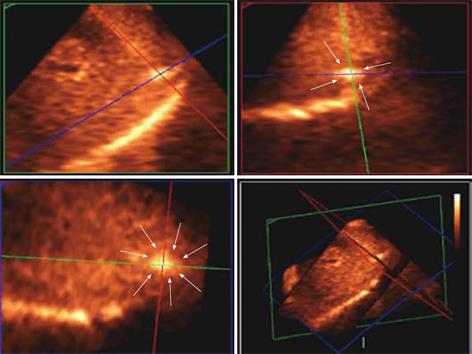
Figure 9 Matrix probe.
3D US showing the hyper-echoic needle electrode tip and its spatial position in the tumour (white arrows delineates the tumour margin) in the subcapsular area and is clearly depicted in 3 orthogonal 2D images confirming the tip being in the center of the tumour.
- Citation: Leen E, Kumar S, Khan SA, Low G, Ong KO, Tait P, Averkiou M. Contrast-enhanced 3D ultrasound in the radiofrequency ablation of liver tumors. World J Gastroenterol 2009; 15(3): 289-299
- URL: https://www.wjgnet.com/1007-9327/full/v15/i3/289.htm
- DOI: https://dx.doi.org/10.3748/wjg.15.289


