Copyright
©2009 The WJG Press and Baishideng.
World J Gastroenterol. Aug 7, 2009; 15(29): 3684-3686
Published online Aug 7, 2009. doi: 10.3748/wjg.15.3684
Published online Aug 7, 2009. doi: 10.3748/wjg.15.3684
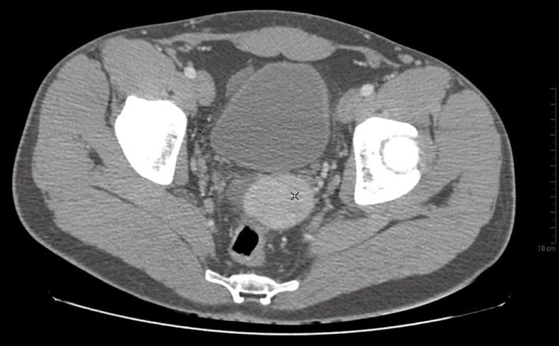
Figure 1 Post-contrast CT scans (after intravenous injection of 130 mL of contrast media, Ultravist, 3 mL/s flow rate).
Lesions are homogeneously hyperdense (92 HU) in the arterial phase (no typical striped enhancement pattern).
- Citation: Garaci FG, Grande M, Villa M, Mancino S, Konda D, Attinà GM, Galatà G, Simonetti G. What is a reliable CT scan for diagnosing splenosis under emergency conditions? World J Gastroenterol 2009; 15(29): 3684-3686
- URL: https://www.wjgnet.com/1007-9327/full/v15/i29/3684.htm
- DOI: https://dx.doi.org/10.3748/wjg.15.3684


