Copyright
©2009 The WJG Press and Baishideng.
World J Gastroenterol. Aug 7, 2009; 15(29): 3670-3675
Published online Aug 7, 2009. doi: 10.3748/wjg.15.3670
Published online Aug 7, 2009. doi: 10.3748/wjg.15.3670
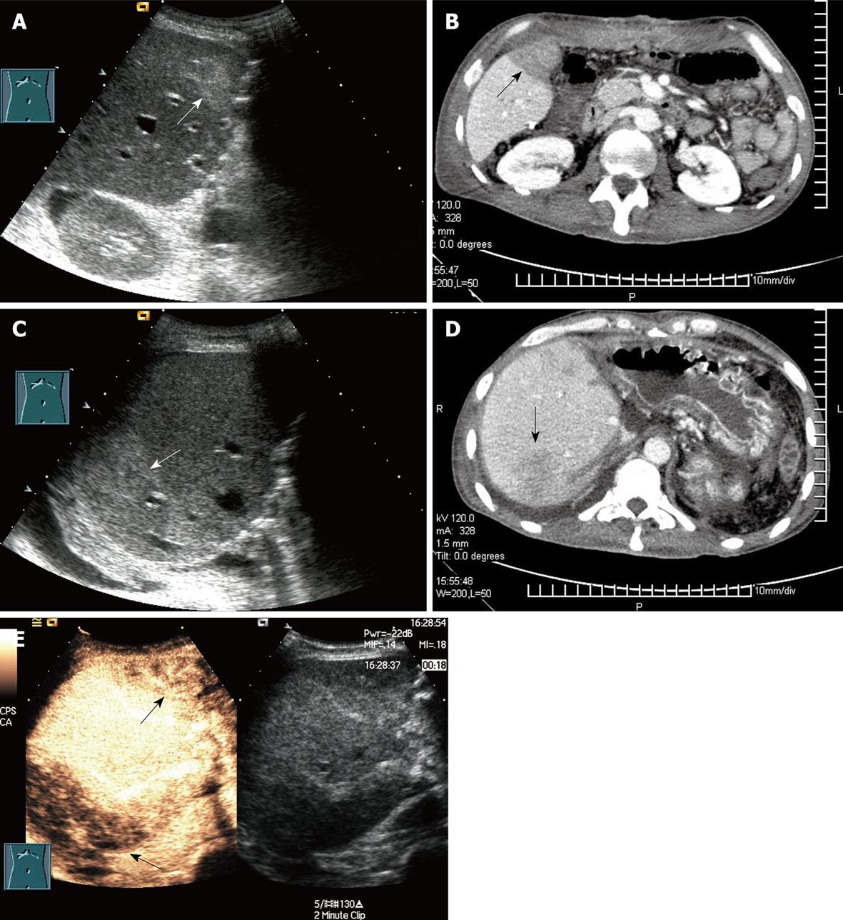
Figure 4 Regional congestion in a 35-year-old man who underwent RLDLT.
A: Longitudinal oblique 2D US scan 1 d after RLDLT revealed regional echogenicity in segment V; B: Contrast-enhanced CT showed poor perfusion (arrow) in segment V 2 d after ultrasound examination; C: Longitudinal oblique 2D US scan revealed regional echogenicity in segment VII; D: Contrast-enhanced CT showed poor perfusion (arrow) in segment VII; E: Longitudinal oblique CEUS scan obtained following the Doppler scan. At CEUS, the poor perfusion area (arrows) was seen as an inhomogenous echo that corresponded to segment V and segment VII. This patient remained asymptomatic and formed collaterals between the tributary of the middle hepatic vein and the right hepatic vein at Doppler ultrasound follow-up.
- Citation: Luo Y, Fan YT, Lu Q, Li B, Wen TF, Zhang ZW. CEUS: A new imaging approach for postoperative vascular complications after right-lobe LDLT. World J Gastroenterol 2009; 15(29): 3670-3675
- URL: https://www.wjgnet.com/1007-9327/full/v15/i29/3670.htm
- DOI: https://dx.doi.org/10.3748/wjg.15.3670


