Copyright
©2009 The WJG Press and Baishideng.
World J Gastroenterol. Jul 28, 2009; 15(28): 3573-3575
Published online Jul 28, 2009. doi: 10.3748/wjg.15.3573
Published online Jul 28, 2009. doi: 10.3748/wjg.15.3573
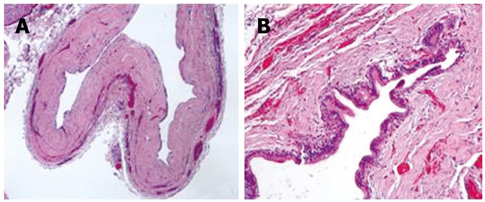
Figure 3 Microscopic view of the sample revealing a multiloculated cystic lesion (A) and a fibrous cyst wall is fibrous lined by epithelium (B).
- Citation: Bartolome MAH, Ruiz SF, Romero IM, Lojo BR, Prieto IR, Alvira LG, Carreño RG, Esteban ML. Biliary cystadenoma. World J Gastroenterol 2009; 15(28): 3573-3575
- URL: https://www.wjgnet.com/1007-9327/full/v15/i28/3573.htm
- DOI: https://dx.doi.org/10.3748/wjg.15.3573


