Copyright
©2009 The WJG Press and Baishideng.
World J Gastroenterol. Jul 28, 2009; 15(28): 3569-3572
Published online Jul 28, 2009. doi: 10.3748/wjg.15.3569
Published online Jul 28, 2009. doi: 10.3748/wjg.15.3569
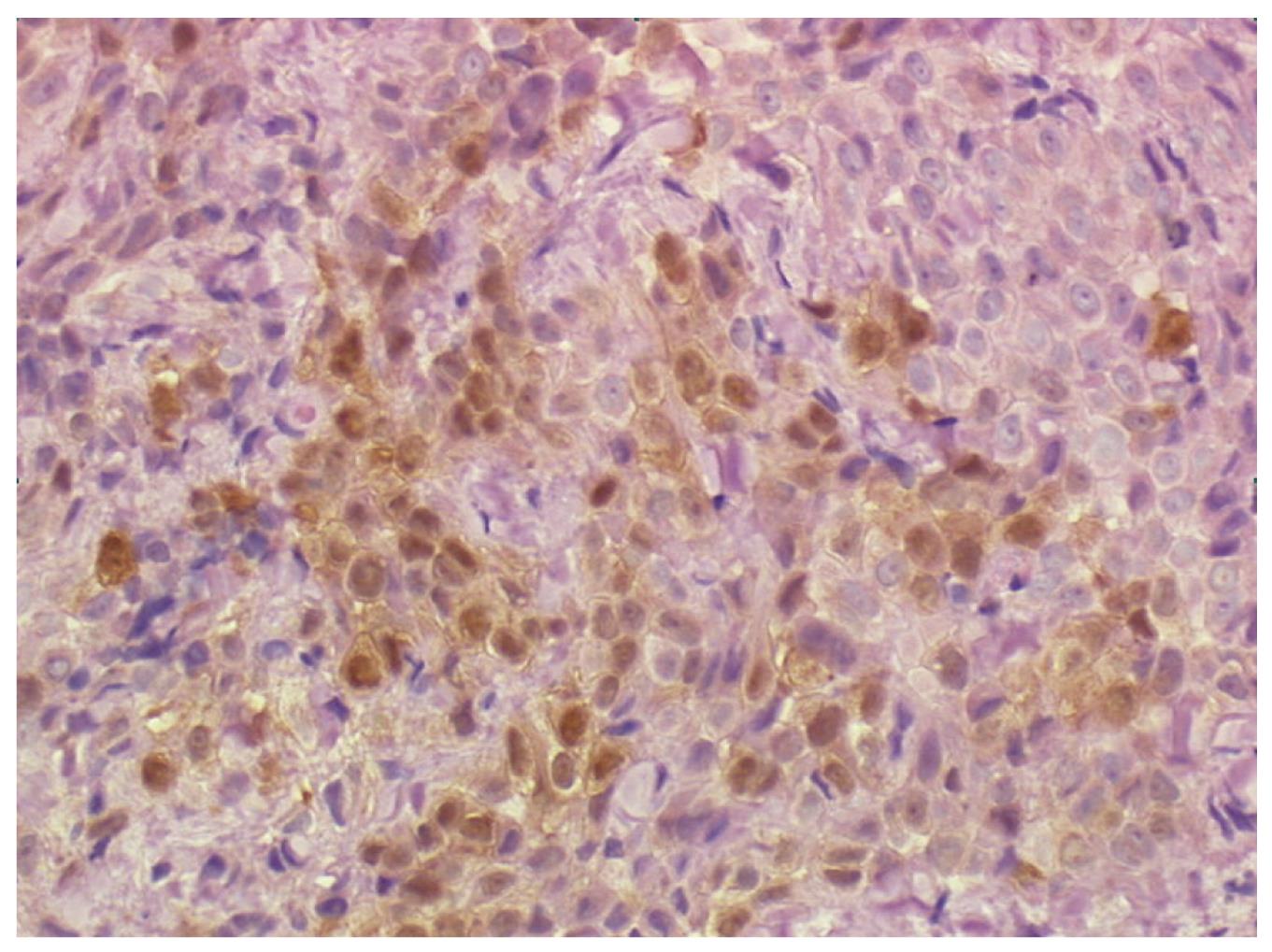
Figure 4 Biopsy of the distal esophageal wall taken at laparoscopy showing a neoplastic cell population consistent with the diagnosis of pleural mesothelioma (epithelioid type).
Immunohistochemistry stains positive for calretinin (40 × HPF).
- Citation: Saino G, Bona D, Nencioni M, Rubino B, Bonavina L. Laparoscopic diagnosis of pleural mesothelioma presenting with pseudoachalasia. World J Gastroenterol 2009; 15(28): 3569-3572
- URL: https://www.wjgnet.com/1007-9327/full/v15/i28/3569.htm
- DOI: https://dx.doi.org/10.3748/wjg.15.3569


