Copyright
©2009 The WJG Press and Baishideng.
World J Gastroenterol. Jul 28, 2009; 15(28): 3480-3485
Published online Jul 28, 2009. doi: 10.3748/wjg.15.3480
Published online Jul 28, 2009. doi: 10.3748/wjg.15.3480
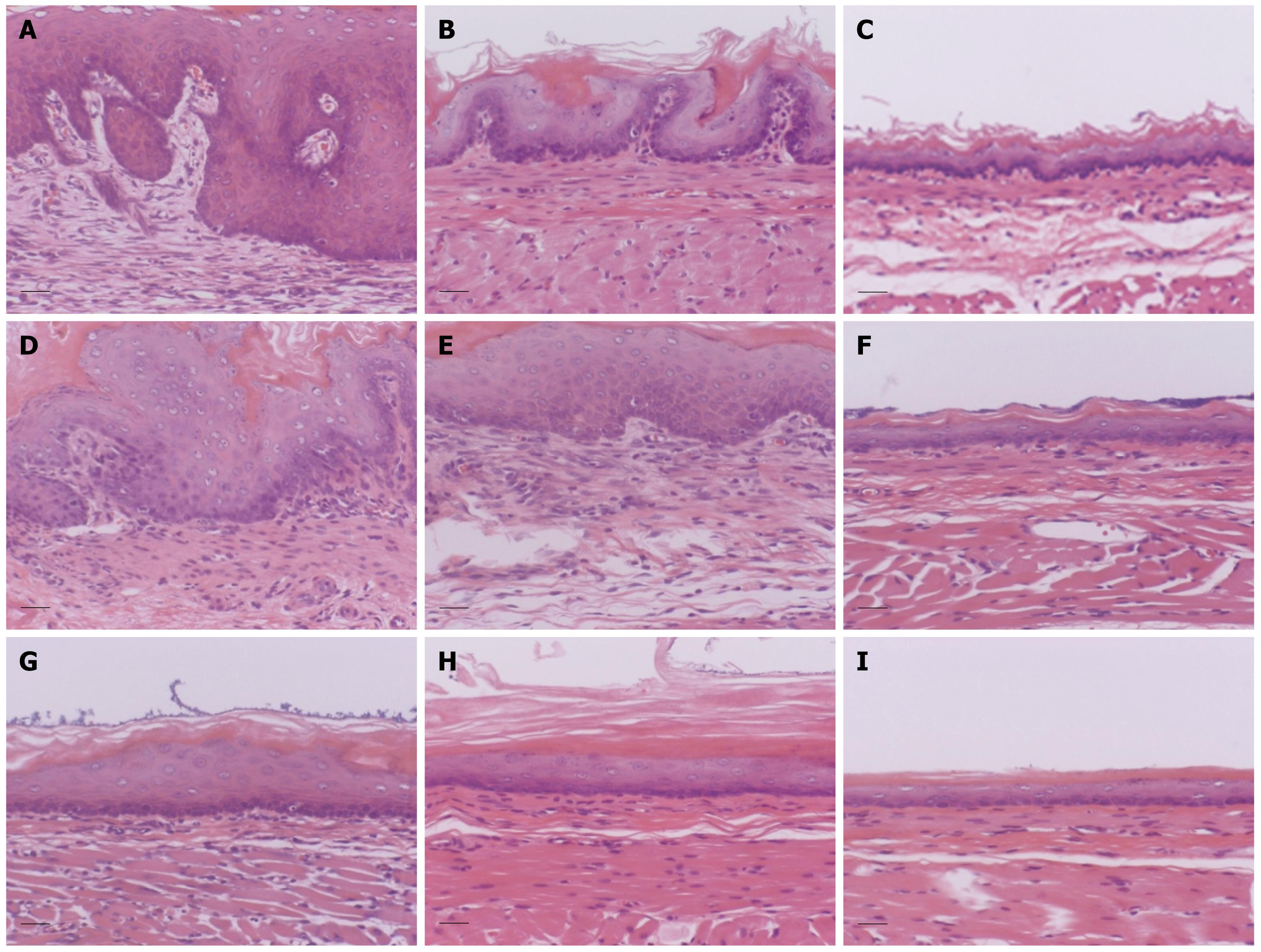
Figure 4 Histological findings in each part of the esophagus in the three groups (HE staining).
Lower esophagus: esophagitis (A), ES (B) and control (C) groups. Middle esophagus: esophagitis (D), ES (E) and control (F) groups. Upper esophagus: esophagitis (G), ES (H) and control (I) groups. In the ES group, the thickness of the epithelium in the lower esophagus was significantly decreased compared with that in the esophagitis group. Scale bar, 30 &mgr;m.
- Citation: Asaoka D, Nagahara A, Oguro M, Izumi Y, Kurosawa A, Osada T, Kawabe M, Hojo M, Otaka M, Watanabe S. Characteristic pathological findings and effects of ecabet sodium in rat reflux esophagitis. World J Gastroenterol 2009; 15(28): 3480-3485
- URL: https://www.wjgnet.com/1007-9327/full/v15/i28/3480.htm
- DOI: https://dx.doi.org/10.3748/wjg.15.3480


