Copyright
©2009 The WJG Press and Baishideng.
World J Gastroenterol. Jul 21, 2009; 15(27): 3440-3444
Published online Jul 21, 2009. doi: 10.3748/wjg.15.3440
Published online Jul 21, 2009. doi: 10.3748/wjg.15.3440
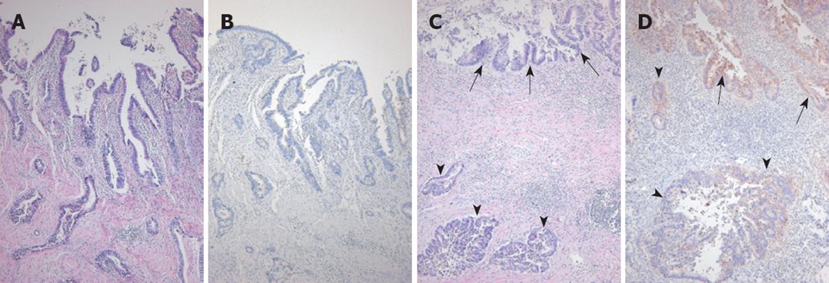
Figure 3 Histopathological finding of hilar bile duct cancer.
A: Histologic section of the gallbladder shows well-differentiated biliary-type adenocarcinoma infiltrating the entire thickness of the gallbladder wall (HE, × 100); B: None of the tumor cells expressed CA19-9 (immunoperoxidase method, × 100); C: Histologic section of the hepatic duct demonstrates severe epithelial dysplasia of the mucosa in the upper field (arrows) and invasive adenocarcinoma in the lower field (arrow heads) (hematoxylin and eosin, × 100); D: Most of the dysplastic epithelial cells in the upper field (arrows) and some of invasive tumor cells in the lower field express cytoplasmic CA19-9 (arrow heads). An invasive tumor gland is noted in the mid-left field (immunoperoxidase method, × 100).
- Citation: Joo HJ, Kim GH, Jeon WJ, Chae HB, Park SM, Youn SJ, Choi JW, Sung R. Metachronous bile duct cancer nine years after resection of gallbladder cancer. World J Gastroenterol 2009; 15(27): 3440-3444
- URL: https://www.wjgnet.com/1007-9327/full/v15/i27/3440.htm
- DOI: https://dx.doi.org/10.3748/wjg.15.3440


