Copyright
©2009 The WJG Press and Baishideng.
World J Gastroenterol. Jul 14, 2009; 15(26): 3315-3318
Published online Jul 14, 2009. doi: 10.3748/wjg.15.3315
Published online Jul 14, 2009. doi: 10.3748/wjg.15.3315
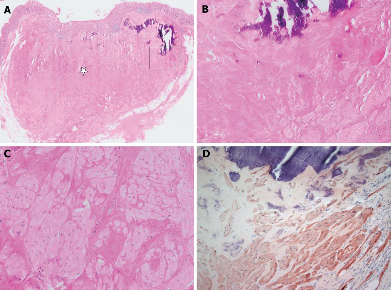
Figure 2 Microscopically and immunohistochemically, the appearance of the tumor was compatible with that of granular cell tumor.
A: The tumor was located mainly between the submucosa and the subserosa. Much of the tumor showed extensive hyalinization (star) and dystrophic calcification. B and C: A high magnification view (square of A) showed dystrophic calcification, hyalinization and some viable tumor cell nests in the peripheral portion of the tumor. The tumor cells were composed of round to polygonal cells with abundant granular eosinophilic cytoplasm. D: Immunohistochemically, tumor cells were reactive for S-100 protein.
- Citation: Hong R, Lim SC. Granular cell tumor of the cecum with extensive hyalinization and calcification: A case report. World J Gastroenterol 2009; 15(26): 3315-3318
- URL: https://www.wjgnet.com/1007-9327/full/v15/i26/3315.htm
- DOI: https://dx.doi.org/10.3748/wjg.15.3315


