Copyright
©2009 The WJG Press and Baishideng.
World J Gastroenterol. Jul 14, 2009; 15(26): 3232-3239
Published online Jul 14, 2009. doi: 10.3748/wjg.15.3232
Published online Jul 14, 2009. doi: 10.3748/wjg.15.3232
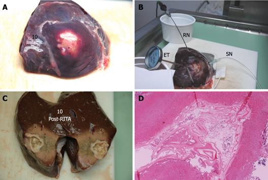
Figure 6 Echinococcus cyst of the liver treated with RTA.
The upper panel shows the surface of the liver before (A) and after (B) RTA. Procedure caused cyst retraction. RITA needle, sentinel needle and external thermometer, still in place, can be seen (B). The left lower panel shows post-RTA section (C) of the cyst. Histology of a paramedian section (right lower panel, HE stain, × 2) shows cyst walls and surrounding liver closest to the cyst are totally necrotic and with granular amorphous aspect (D). RN: RITA needle; SN: Sentinel needle; ET: External thermometer.
-
Citation: Lamonaca V, Virga A, Minervini MI, Di Stefano R, Provenzani A, Tagliareni P, Fleres G, Luca A, Vizzini G, Palazzo U, Gridelli B. Cystic echinococcosis of the liver and lung treated by radiofrequency thermal ablation: An
ex-vivo pilot experimental study in animal models. World J Gastroenterol 2009; 15(26): 3232-3239 - URL: https://www.wjgnet.com/1007-9327/full/v15/i26/3232.htm
- DOI: https://dx.doi.org/10.3748/wjg.15.3232


