Copyright
©2009 The WJG Press and Baishideng.
World J Gastroenterol. Jul 14, 2009; 15(26): 3232-3239
Published online Jul 14, 2009. doi: 10.3748/wjg.15.3232
Published online Jul 14, 2009. doi: 10.3748/wjg.15.3232
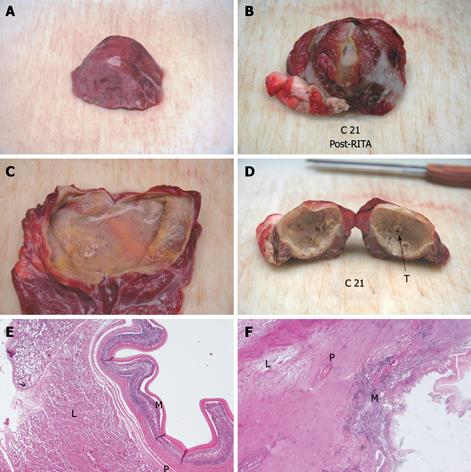
Figure 4 Hydatid cyst of the lung.
Untreated (left) and treated (right) cysts are compared. External surface (upper panel), findings after sectioning (middle panel), and 4 × HE stained histology (lower panel) are shown. Untreated cyst shows normal surface, flaccidity after sectioning, thin and translucid endocyst and well preserved cyst layers at histology. Treated cyst looks dehydrated, rigid, has a thick and papyraceous-like endocyst and is totally necrotic at histology. T: RITA-needle through; M: Germinal membrane; P: Pericystium; L: Lung parenchyma.
-
Citation: Lamonaca V, Virga A, Minervini MI, Di Stefano R, Provenzani A, Tagliareni P, Fleres G, Luca A, Vizzini G, Palazzo U, Gridelli B. Cystic echinococcosis of the liver and lung treated by radiofrequency thermal ablation: An
ex-vivo pilot experimental study in animal models. World J Gastroenterol 2009; 15(26): 3232-3239 - URL: https://www.wjgnet.com/1007-9327/full/v15/i26/3232.htm
- DOI: https://dx.doi.org/10.3748/wjg.15.3232


