Copyright
©2009 The WJG Press and Baishideng.
World J Gastroenterol. Jun 21, 2009; 15(23): 2930-2932
Published online Jun 21, 2009. doi: 10.3748/wjg.15.2930
Published online Jun 21, 2009. doi: 10.3748/wjg.15.2930
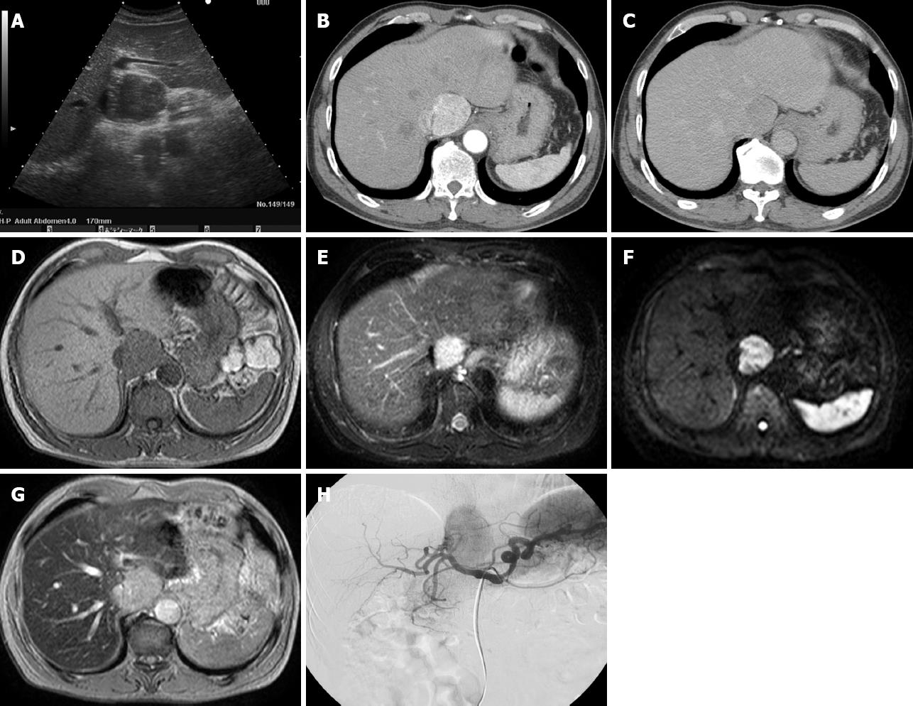
Figure 1 Images.
A: The tumor was hypoechoic on ultrasonography, measuring 4.2 cm × 4.0 cm; B-C: Enhanced computed tomography showed a tumor with early-phase hyperattenuation and late-phase hypoattenuation; D-F: Magnetic resonance imaging showed a tumor with hypointensity on T1-weighted, hyperintensity on T2-weighted images and hyperintensity on diffusion weighted images; G: The tumor did not absorb iron on superparamagnetic iron oxide-enhanced MRI; H: On angiography, the tumor was shown as a circumscribed hypervascular mass.
- Citation: Takahara M, Miyake Y, Matsumoto K, Kawai D, Kaji E, Toyokawa T, Nakatsu M, Ando M, Hirohata M. A case of hepatic angiomyolipoma difficult to distinguish from hepatocellular carcinoma. World J Gastroenterol 2009; 15(23): 2930-2932
- URL: https://www.wjgnet.com/1007-9327/full/v15/i23/2930.htm
- DOI: https://dx.doi.org/10.3748/wjg.15.2930


