Copyright
©2009 The WJG Press and Baishideng.
World J Gastroenterol. Jun 21, 2009; 15(23): 2927-2929
Published online Jun 21, 2009. doi: 10.3748/wjg.15.2927
Published online Jun 21, 2009. doi: 10.3748/wjg.15.2927
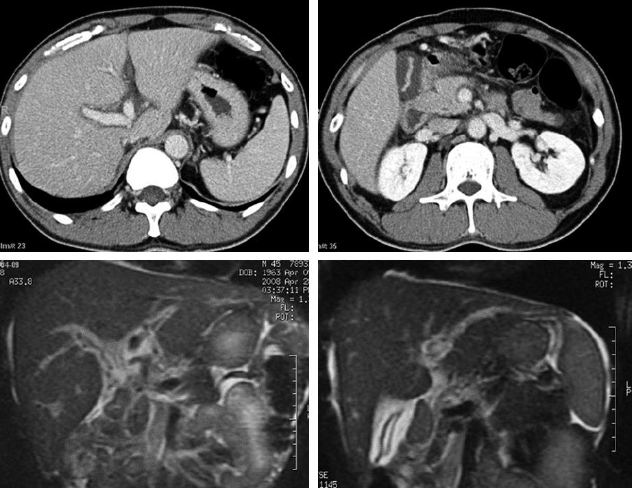
Figure 1 Abdominal CTs and MRIs on admission.
Abdominal CTs showing normal liver shape and architecture, normal intra-and extrahepatic bile ducts, and a severely swollen gallbladder (top); MRIs showing a normal liver, bile duct, an edematous, thickened gallbladder wall, and periportal edema (bottom).
- Citation: Ohwada S, Kobayashi I, Harasawa N, Tsuda K, Inui Y. Severe acute cholestatic hepatitis of unknown etiology successfully treated with the Chinese herbal medicine Inchinko-to (TJ-135). World J Gastroenterol 2009; 15(23): 2927-2929
- URL: https://www.wjgnet.com/1007-9327/full/v15/i23/2927.htm
- DOI: https://dx.doi.org/10.3748/wjg.15.2927


