Copyright
©2009 The WJG Press and Baishideng.
World J Gastroenterol. Jun 21, 2009; 15(23): 2923-2926
Published online Jun 21, 2009. doi: 10.3748/wjg.15.2923
Published online Jun 21, 2009. doi: 10.3748/wjg.15.2923
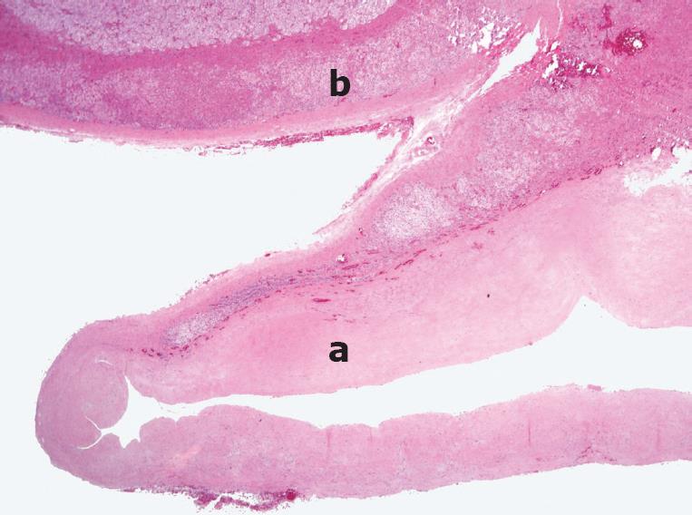
Figure 5 Histopathology showing an adrenal pseudocyst (a) and adrenal tissue remnants on the upper aspect (b).
The cyst wall is composed of a thick layer of hyalinized connective tissue without an epithelial or endothelial lining (a). HE staining (Original magnification, × 40).
- Citation: Kim BS, Joo SH, Choi SI, Song JY. Laparoscopic resection of an adrenal pseudocyst mimicking a retroperitoneal mucinous cystic neoplasm. World J Gastroenterol 2009; 15(23): 2923-2926
- URL: https://www.wjgnet.com/1007-9327/full/v15/i23/2923.htm
- DOI: https://dx.doi.org/10.3748/wjg.15.2923


