Copyright
©2009 The WJG Press and Baishideng.
World J Gastroenterol. Jun 14, 2009; 15(22): 2739-2747
Published online Jun 14, 2009. doi: 10.3748/wjg.15.2739
Published online Jun 14, 2009. doi: 10.3748/wjg.15.2739
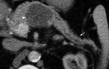
Figure 6 Axial MDCT image from a 64-year-old woman with an incidentally discovered cystic lesion in the body of the pancreas.
A large serous cystadenoma (arrow) present in the body of pancreas displays microcystic morphology and fine lobulations with a central scar. Atrophy of the pancreatic parenchyma distal to the site of the lesion is also present (small arrow).
- Citation: Shah AA, Sainani NI, Ramesh AK, Shah ZK, Deshpande V, Hahn PF, Sahani DV. Predictive value of multi-detector computed tomography for accurate diagnosis of serous cystadenoma: Radiologic-pathologic correlation. World J Gastroenterol 2009; 15(22): 2739-2747
- URL: https://www.wjgnet.com/1007-9327/full/v15/i22/2739.htm
- DOI: https://dx.doi.org/10.3748/wjg.15.2739


