Copyright
©2009 The WJG Press and Baishideng.
World J Gastroenterol. Jun 14, 2009; 15(22): 2739-2747
Published online Jun 14, 2009. doi: 10.3748/wjg.15.2739
Published online Jun 14, 2009. doi: 10.3748/wjg.15.2739
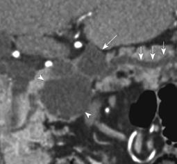
Figure 5 Coronal reformatted MDCT image from an asymptomatic 90-year-old woman, who had a 5 cm lesion in the head of the pancreas which was removed by Whipple procedure.
The image shows an oligocystic SCA (arrow heads) in the head of pancreas which was mistaken for a mucinous lesion due to an associated side branch IPMN adjacent to it (white arrow). The two lesions were interpreted as a single multiloculated side branch mucinous lesion. There is mild upstream dilatation of the visualized pancreatic duct (small white arrows).
- Citation: Shah AA, Sainani NI, Ramesh AK, Shah ZK, Deshpande V, Hahn PF, Sahani DV. Predictive value of multi-detector computed tomography for accurate diagnosis of serous cystadenoma: Radiologic-pathologic correlation. World J Gastroenterol 2009; 15(22): 2739-2747
- URL: https://www.wjgnet.com/1007-9327/full/v15/i22/2739.htm
- DOI: https://dx.doi.org/10.3748/wjg.15.2739


