Copyright
©2009 The WJG Press and Baishideng.
World J Gastroenterol. Jun 14, 2009; 15(22): 2739-2747
Published online Jun 14, 2009. doi: 10.3748/wjg.15.2739
Published online Jun 14, 2009. doi: 10.3748/wjg.15.2739
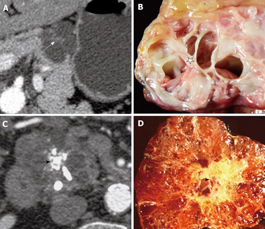
Figure 3 Images from 2 different patients with central scar.
A: Axial MDCT image from a 46-year-old woman with a macrocystic SCA with a central scar (white arrow); B: Gross pathological specimen of the same patient also reveals the macrocystic pattern with the septa converging on a central scar (star); C: Axial MDCT image from a 66-year-old male who had a history of chronic pancreatitis, reveals a large lobulated microcystic lesion with central stellate calcification (black arrow). A Whipple procedure was done to remove a 4 cm mass from the head of the pancreas. Corresponding gross pathological image of the microcystic SCA with calcification (black arrow) in the central scar (D).
- Citation: Shah AA, Sainani NI, Ramesh AK, Shah ZK, Deshpande V, Hahn PF, Sahani DV. Predictive value of multi-detector computed tomography for accurate diagnosis of serous cystadenoma: Radiologic-pathologic correlation. World J Gastroenterol 2009; 15(22): 2739-2747
- URL: https://www.wjgnet.com/1007-9327/full/v15/i22/2739.htm
- DOI: https://dx.doi.org/10.3748/wjg.15.2739


