Copyright
©2009 The WJG Press and Baishideng.
World J Gastroenterol. Jun 14, 2009; 15(22): 2739-2747
Published online Jun 14, 2009. doi: 10.3748/wjg.15.2739
Published online Jun 14, 2009. doi: 10.3748/wjg.15.2739
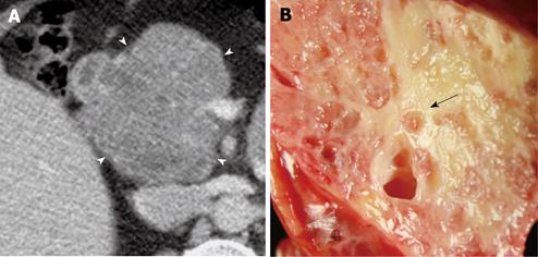
Figure 1 A 64-year-old woman who was incidentally found to have a mass in the head of the pancreas and subsequently underwent a Whipple procedure for removal.
A: Lobulated (arrow heads) microcystic serous cystadenoma (SCA) with characteristic honeycomb appearance is seen on axial MDCT image (1.25 mm); B: Gross pathological specimen of the lesion reveals cluster of microcysts with a sponge pattern. A central scar is appreciated on the pathological image (black arrow), which is subtle and difficult to appreciate on the CT image.
- Citation: Shah AA, Sainani NI, Ramesh AK, Shah ZK, Deshpande V, Hahn PF, Sahani DV. Predictive value of multi-detector computed tomography for accurate diagnosis of serous cystadenoma: Radiologic-pathologic correlation. World J Gastroenterol 2009; 15(22): 2739-2747
- URL: https://www.wjgnet.com/1007-9327/full/v15/i22/2739.htm
- DOI: https://dx.doi.org/10.3748/wjg.15.2739


