Copyright
©2009 The WJG Press and Baishideng.
World J Gastroenterol. Jun 7, 2009; 15(21): 2657-2664
Published online Jun 7, 2009. doi: 10.3748/wjg.15.2657
Published online Jun 7, 2009. doi: 10.3748/wjg.15.2657
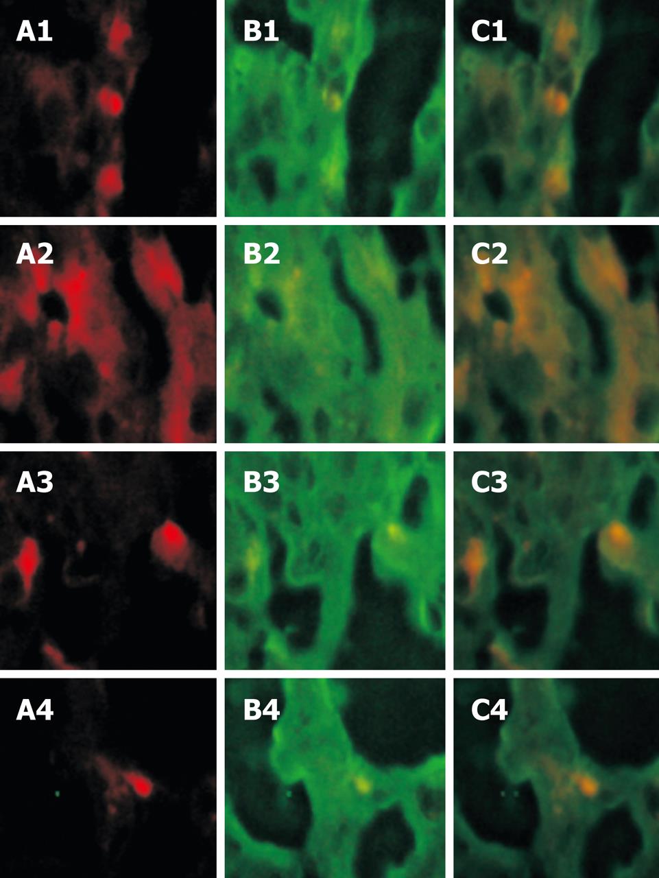
Figure 3 Confocal microscopy shows red fluorescence of cell location and green fluorescence of albumin.
The red fluorescence cells could be found in liver tissue of recipient mice, suggesting that PHK-26 positive cells can emerge out of the red fluorescence (A1-A4). The albumin expressed in hepatocytes showed green fluorescence (B1-B4). After the red and green fluorescence cells were located, yellow cells were found in a suitable location (C1-C4).
- Citation: Jin SZ, Meng XW, Han MZ, Sun X, Sun LY, Liu BR. Stromal cell derived factor-1 enhances bone marrow mononuclear cell migration in mice with acute liver failure. World J Gastroenterol 2009; 15(21): 2657-2664
- URL: https://www.wjgnet.com/1007-9327/full/v15/i21/2657.htm
- DOI: https://dx.doi.org/10.3748/wjg.15.2657


