Copyright
©2009 The WJG Press and Baishideng.
World J Gastroenterol. Jun 7, 2009; 15(21): 2609-2616
Published online Jun 7, 2009. doi: 10.3748/wjg.15.2609
Published online Jun 7, 2009. doi: 10.3748/wjg.15.2609
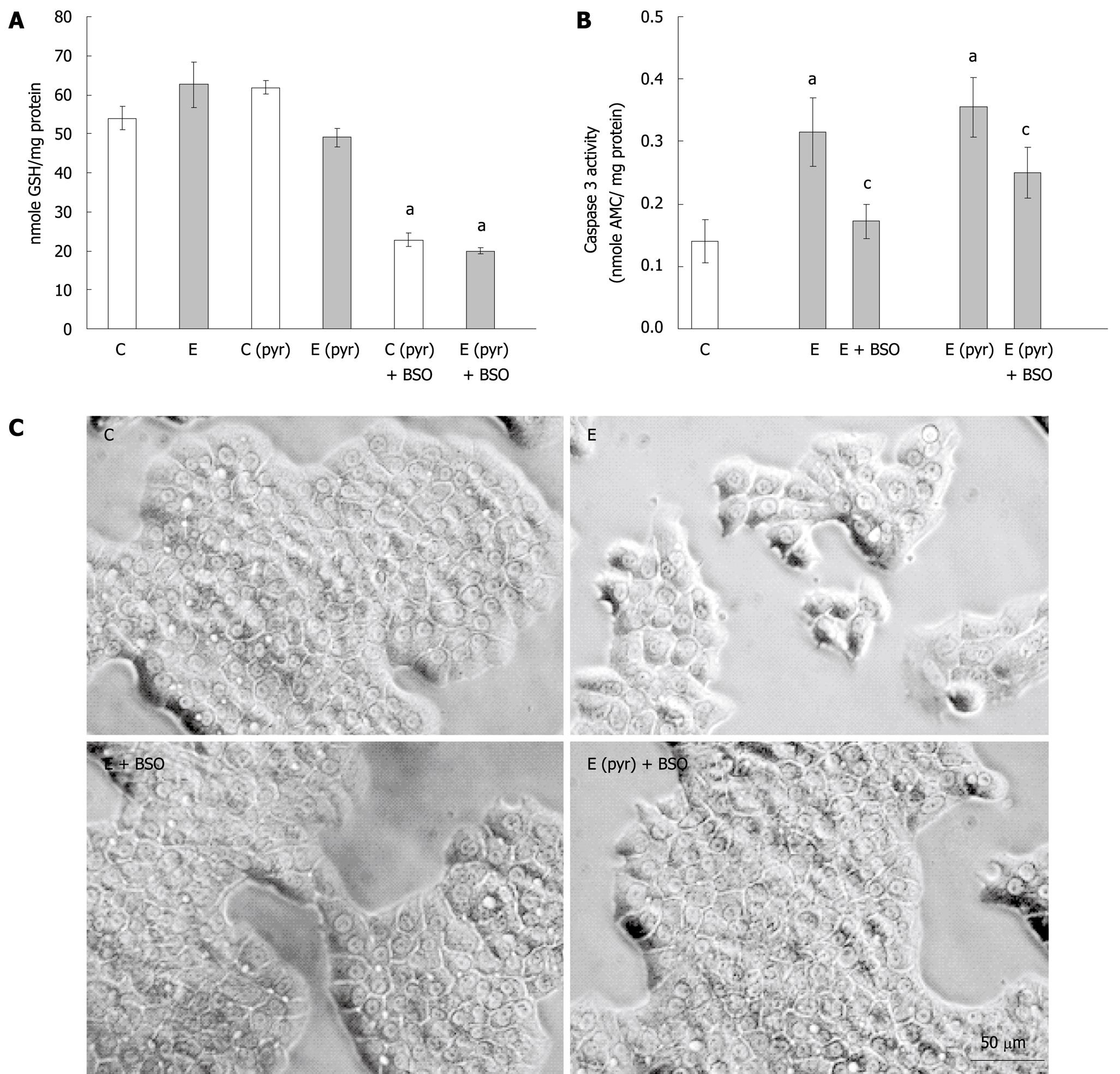
Figure 3 The effect of glutathione depletion on the induction of caspase activation in ethanol-treated WIF-B cells.
WIF-B cultures were treated in the presence or absence of ethanol (25 mmol/L) for 48 h after pretreatment with (C pyr and E pyr) or without (C and E) pyrazole. Glutathione was depleted by the inclusion of BSO in the culture media when indicated (+ BSO). A: The amount of reduced glutathione (GSH) was detected as described in “Material and Methods”; B: Measure of apoptosis in WIF-B cells following glutathione depletion. Cell lysates were assayed for caspase-3 activity and the release of fluorescent product was detected and expressed for five independent experiments; C: Phase contrast images of the treated WIF-B cultures. aP < 0.05 vs control; cP < 0.05 vs ethanol-treated.
- Citation: McVicker BL, Tuma PL, Kharbanda KK, Lee SM, Tuma DJ. Relationship between oxidative stress and hepatic glutathione levels in ethanol-mediated apoptosis of polarized hepatic cells. World J Gastroenterol 2009; 15(21): 2609-2616
- URL: https://www.wjgnet.com/1007-9327/full/v15/i21/2609.htm
- DOI: https://dx.doi.org/10.3748/wjg.15.2609


