Copyright
©2009 The WJG Press and Baishideng.
World J Gastroenterol. Jan 14, 2009; 15(2): 198-203
Published online Jan 14, 2009. doi: 10.3748/wjg.15.198
Published online Jan 14, 2009. doi: 10.3748/wjg.15.198
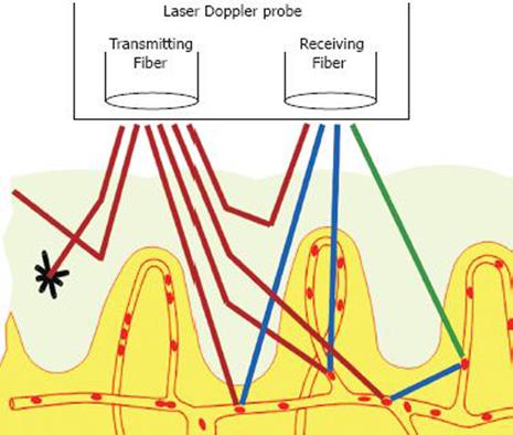
Figure 1 A schematic depiction of laser Doppler perfusion monitoring showing the probe with its emitting fibre bundle which applies monochro-matic laser light to the tissue, and its receiving fibre bundle which returns reflected light for analysis.
The light that has undergone a doppler shift due to moving blood cells in the tissues reflects the microcirculatory perfusion at a given time. Reproduced by permission of Perimed AB.
- Citation: Hoff DAL, Gregersen H, Hatlebakk JG. Mucosal blood flow measurements using laser Doppler perfusion monitoring. World J Gastroenterol 2009; 15(2): 198-203
- URL: https://www.wjgnet.com/1007-9327/full/v15/i2/198.htm
- DOI: https://dx.doi.org/10.3748/wjg.15.198


