Copyright
©2009 The WJG Press and Baishideng.
World J Gastroenterol. Jan 14, 2009; 15(2): 192-197
Published online Jan 14, 2009. doi: 10.3748/wjg.15.192
Published online Jan 14, 2009. doi: 10.3748/wjg.15.192
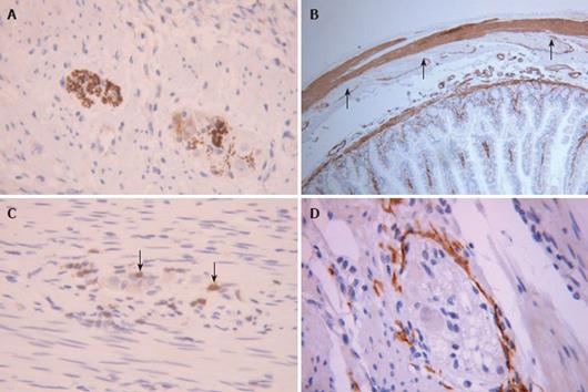
Figure 3 Immunohistochemistry using antibodies to.
A: Neuron specific enolase allowing clear visualisation of myenteric ganglia, neuronal number and size; B: Smooth muscle alpha actin showing absent staining in the circular muscle layer of the jejunum (arrows) in a patient with enteric dysmotility; C: CD3 showing small numbers of periganglionic T lymphocytes (arrows) in numbers that most would deem abnormal and indicative of ganglionitis; D: CD117 staining showing normal myenteric plexus interstitial cells of Cajal (ICC-MP). (Original magnification × 40-100).
- Citation: Knowles CH, Martin JE. New techniques in the tissue diagnosis of gastrointestinal neuromuscular diseases. World J Gastroenterol 2009; 15(2): 192-197
- URL: https://www.wjgnet.com/1007-9327/full/v15/i2/192.htm
- DOI: https://dx.doi.org/10.3748/wjg.15.192


