Copyright
©2009 The WJG Press and Baishideng.
World J Gastroenterol. Jan 14, 2009; 15(2): 192-197
Published online Jan 14, 2009. doi: 10.3748/wjg.15.192
Published online Jan 14, 2009. doi: 10.3748/wjg.15.192
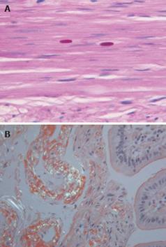
Figure 2 Tinctorial stains used in GI neuromuscular histopathology.
A: Periodic acid Schiff staining showing polyglucosan bodies in a patient with intestinal pseudo-obstruction; B: Bifringence from amyloid visualised by Congo red staining (× 25-40).
- Citation: Knowles CH, Martin JE. New techniques in the tissue diagnosis of gastrointestinal neuromuscular diseases. World J Gastroenterol 2009; 15(2): 192-197
- URL: https://www.wjgnet.com/1007-9327/full/v15/i2/192.htm
- DOI: https://dx.doi.org/10.3748/wjg.15.192


