Copyright
©2009 The WJG Press and Baishideng.
World J Gastroenterol. Jan 14, 2009; 15(2): 182-191
Published online Jan 14, 2009. doi: 10.3748/wjg.15.182
Published online Jan 14, 2009. doi: 10.3748/wjg.15.182
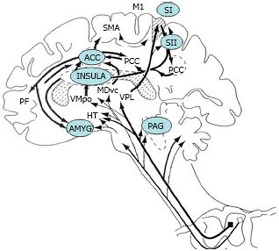
Figure 2 The subcortical and cortical structures that have been shown to be activated in response to visceral pain.
PAG: Periaqueductal grey; PB: Parabrachial nucleus of the dorsolateral pons; VMpo: Ventromedial part of the posterior thalamic nuclear complex; MDvc: Ventrocaudal part of the medial thalamic dorsal nucleus; VPL: Ventroposterior lateral thalamic nucleus; ACC: Anterior cingulate cortex; PCC: Posterior cingulate cortex; HT: Hypothalamus; S1, S2: First and second somatosensory cortical areas, respectively; PPC: Posterior parietal complex; SMA: Supplementary motor area; AMYG: Amygdala; PF: Prefrontal cortex; M1: Motor cortex. (Adapted from Price DD. Psychological and neural mechanisms of the affective dimension of pain. Science 2000; 288: 1769-1772).
- Citation: Sharma A, Lelic D, Brock C, Paine P, Aziz Q. New technologies to investigate the brain-gut axis. World J Gastroenterol 2009; 15(2): 182-191
- URL: https://www.wjgnet.com/1007-9327/full/v15/i2/182.htm
- DOI: https://dx.doi.org/10.3748/wjg.15.182


