Copyright
©2009 The WJG Press and Baishideng.
World J Gastroenterol. Jan 14, 2009; 15(2): 182-191
Published online Jan 14, 2009. doi: 10.3748/wjg.15.182
Published online Jan 14, 2009. doi: 10.3748/wjg.15.182
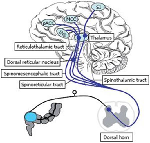
Figure 1 The principal visceral projections from the spinal cord to subcortial and cortical structures (blue lines).
The spinothalamic tract terminates in the medial and posterior thalamus. Thalamocortical fibres then project to the primary somatosensory cortex. The spinoreticular tract terminates in the reticular formation to the medial thalamus. The spinomesencephalic tract projects to various regions in the brainstem, including the periaqueductal grey, locus coeruleus, and dorsal reticular nucleus in the medulla. Thalamocortical projections from the medial thalamus project to the cingulate cortex and insula which are involved in processing noxious visceral and somatic information. The brain regions innervated by these pathways that respond to painful visceral stimuli include the thalamus, insula, amygdala and anterior cingulate cortex (ACC). The ACC is comprised of two components, the perigenual ACC (pACC) involved in affect and the mid cingulate cortex (MCC) with behavioural response modification. Other pathways for transmission of noxious visceral stimuli (such as the dorsal column pathway), exist, but are not shown here.
- Citation: Sharma A, Lelic D, Brock C, Paine P, Aziz Q. New technologies to investigate the brain-gut axis. World J Gastroenterol 2009; 15(2): 182-191
- URL: https://www.wjgnet.com/1007-9327/full/v15/i2/182.htm
- DOI: https://dx.doi.org/10.3748/wjg.15.182


