Copyright
©2009 The WJG Press and Baishideng.
World J Gastroenterol. May 21, 2009; 15(19): 2389-2394
Published online May 21, 2009. doi: 10.3748/wjg.15.2389
Published online May 21, 2009. doi: 10.3748/wjg.15.2389
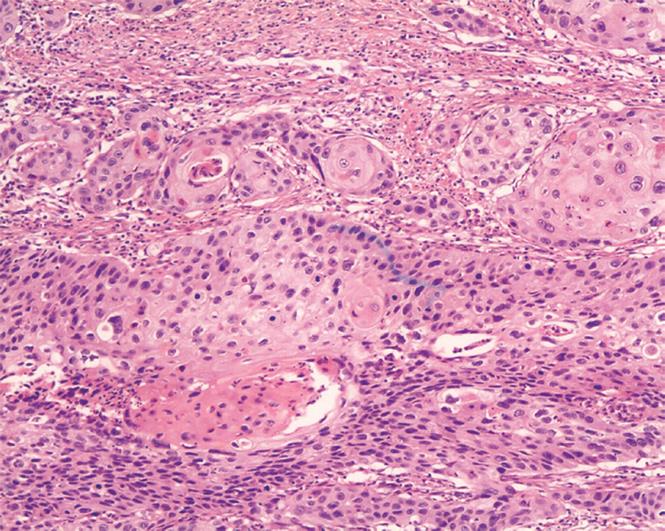
Figure 2 Staining of ESCC tissues.
The tumor cells of cancerous tissues were stained as violet in the nucleus and pink in the cytoplasm (× 100).
- Citation: He XT, Cao XF, Ji L, Zhu B, Lv J, Wang DD, Lu PH, Cui HG. Association between Bmi1 and clinicopathological status of esophageal squamous cell carcinoma. World J Gastroenterol 2009; 15(19): 2389-2394
- URL: https://www.wjgnet.com/1007-9327/full/v15/i19/2389.htm
- DOI: https://dx.doi.org/10.3748/wjg.15.2389


