Copyright
©2009 The WJG Press and Baishideng.
World J Gastroenterol. May 14, 2009; 15(18): 2287-2289
Published online May 14, 2009. doi: 10.3748/wjg.15.2287
Published online May 14, 2009. doi: 10.3748/wjg.15.2287
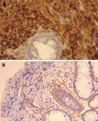
Figure 2 Immunohistochemical staining.
The lesion consists of a pure population of Schwann cells, as shown by the diffuse immunoreactivity for the S-100 protein (A). Only scattered myoepithelial cells and vascular structures were highlighted by the immunostaining for α-smooth muscle actin (B).
- Citation: Pasquini P, Baiocchini A, Falasca L, Annibali D, Gimbo G, Pace F, Nonno FD. Mucosal Schwann cell “Hamartoma”: A new entity? World J Gastroenterol 2009; 15(18): 2287-2289
- URL: https://www.wjgnet.com/1007-9327/full/v15/i18/2287.htm
- DOI: https://dx.doi.org/10.3748/wjg.15.2287


