Copyright
©2009 The WJG Press and Baishideng.
World J Gastroenterol. May 14, 2009; 15(18): 2280-2282
Published online May 14, 2009. doi: 10.3748/wjg.15.2280
Published online May 14, 2009. doi: 10.3748/wjg.15.2280
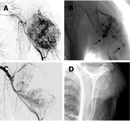
Figure 3 Catheter angiography of the tumor before (A, B) and after embolization (C), X-Ray after final treatment with amputation of the left upper extremity (D).
A: Catheter angiography of the axillary artery reveal a round hypervascular tumor; B: Selective angiography of a tumor feeding vessel as side of application of Bead Block (size 300-500 &mgr;m, Terumo Europe, Leuven, Belgium) for embolization; C: After embolization of main parts of the tumor, at this status no areas of bleeding detectable but small parts of the tumor still perfused; D: Final treatment with amputation of the left upper extremity.
- Citation: Hansch A, Neumann R, Pfeil A, Marintchev I, Pfleiderer S, Gajda M, Kaiser WA. Embolization of an unusual metastatic site of hepatocellular carcinoma in the humerus. World J Gastroenterol 2009; 15(18): 2280-2282
- URL: https://www.wjgnet.com/1007-9327/full/v15/i18/2280.htm
- DOI: https://dx.doi.org/10.3748/wjg.15.2280


