Copyright
©2009 The WJG Press and Baishideng.
World J Gastroenterol. May 14, 2009; 15(18): 2280-2282
Published online May 14, 2009. doi: 10.3748/wjg.15.2280
Published online May 14, 2009. doi: 10.3748/wjg.15.2280
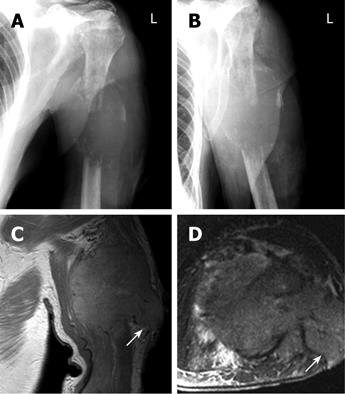
Figure 2 X-Ray (A, B) and MR imaging (C, D) of HCC metastasis at the humerus.
A and B: Destructive lesion in the left humerus associated with a bulging soft tissue component, 5.5 cm × 4 cm in dimension; C: Coronal noncontrast PD-weighted (TR/TE, 1920/12) image demonstrating a soft tissue tumor (coronal dimension 6.2 cm × 5 cm) with complete destruction of the humerus, the tumor reached the surface with ulceration (arrow); D: Axial noncontrast TIRM sequences (TR/TE, 7400/92) show the edematous tumor in the centre of the extremity and the lateral tumor branch to the surface (arrow). In this region, a biopsy was taken which was followed by massive hemorrhage.
- Citation: Hansch A, Neumann R, Pfeil A, Marintchev I, Pfleiderer S, Gajda M, Kaiser WA. Embolization of an unusual metastatic site of hepatocellular carcinoma in the humerus. World J Gastroenterol 2009; 15(18): 2280-2282
- URL: https://www.wjgnet.com/1007-9327/full/v15/i18/2280.htm
- DOI: https://dx.doi.org/10.3748/wjg.15.2280


