Copyright
©2009 The WJG Press and Baishideng.
World J Gastroenterol. May 14, 2009; 15(18): 2277-2279
Published online May 14, 2009. doi: 10.3748/wjg.15.2277
Published online May 14, 2009. doi: 10.3748/wjg.15.2277
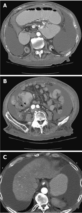
Figure 2 CT scan of the abdomen.
showing stones in the left kidney (A, arrow), pneumatosis intestinalis (B, arrow) and shrunken nodular liver with abdominal ascites (C).
- Citation: Singh D, Laya AS, Clarkston WK, Allen MJ. Jejunoileal bypass: A surgery of the past and a review of its complications. World J Gastroenterol 2009; 15(18): 2277-2279
- URL: https://www.wjgnet.com/1007-9327/full/v15/i18/2277.htm
- DOI: https://dx.doi.org/10.3748/wjg.15.2277


