Copyright
©2009 The WJG Press and Baishideng.
World J Gastroenterol. May 14, 2009; 15(18): 2245-2251
Published online May 14, 2009. doi: 10.3748/wjg.15.2245
Published online May 14, 2009. doi: 10.3748/wjg.15.2245
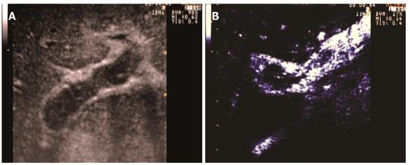
Figure 4 Malignant mosaic thrombus.
A: Sonography scan reveals isoechoic area within portal lumen representing thrombus; B: Contrast enhanced sonography scan during late arterial phase reveals thrombus as predominantly enhancing area, indicative of arterial neovascularization (malignant thrombosis) with some non-enhancing areas of the thrombus (mosaic pattern).
-
Citation: Sorrentino P, D’Angelo S, Tarantino L, Ferbo U, Bracigliano A, Vecchione R. Contrast-enhanced sonography
versus biopsy for the differential diagnosis of thrombosis in hepatocellular carcinoma patients. World J Gastroenterol 2009; 15(18): 2245-2251 - URL: https://www.wjgnet.com/1007-9327/full/v15/i18/2245.htm
- DOI: https://dx.doi.org/10.3748/wjg.15.2245


