Copyright
©2009 The WJG Press and Baishideng.
World J Gastroenterol. May 14, 2009; 15(18): 2245-2251
Published online May 14, 2009. doi: 10.3748/wjg.15.2245
Published online May 14, 2009. doi: 10.3748/wjg.15.2245
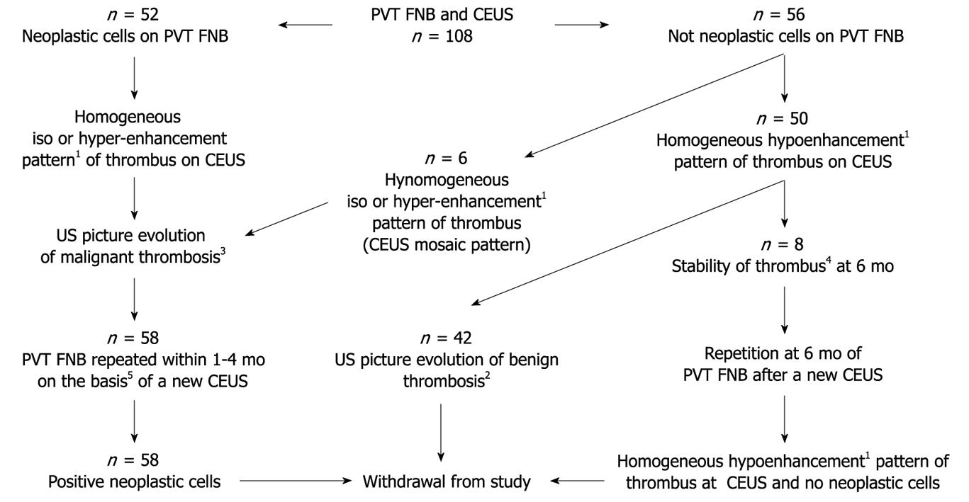
Figure 2 Summary of combined test results.
1Iso, hyper, or hypoenhancement pattern of thrombus compared to the surrounding parenchyma; 2Reference standard of benign thrombosis is a US evidence of evolving thrombus: no increase in size or distribution with vessel wall preservation or recanalization/shrinkage, or disappearance of a PVT within the 6 mo of follow-up were accepted as evidence of a benign portal vein thrombus; 3Us image of evolution, indicating malignant thrombosis: increase in size with infiltration of perivascular parenchyma and interruption of vessel wall was US features of malignant thrombosis; 4No change in thrombus image and in the diameter of the segment of vein involved at 6 mo of follow-up; 5PVT FNB were repeated guiding the needle to the thrombus territories with enhancing pattern.
-
Citation: Sorrentino P, D’Angelo S, Tarantino L, Ferbo U, Bracigliano A, Vecchione R. Contrast-enhanced sonography
versus biopsy for the differential diagnosis of thrombosis in hepatocellular carcinoma patients. World J Gastroenterol 2009; 15(18): 2245-2251 - URL: https://www.wjgnet.com/1007-9327/full/v15/i18/2245.htm
- DOI: https://dx.doi.org/10.3748/wjg.15.2245


