Copyright
©2009 The WJG Press and Baishideng.
World J Gastroenterol. May 14, 2009; 15(18): 2214-2219
Published online May 14, 2009. doi: 10.3748/wjg.15.2214
Published online May 14, 2009. doi: 10.3748/wjg.15.2214
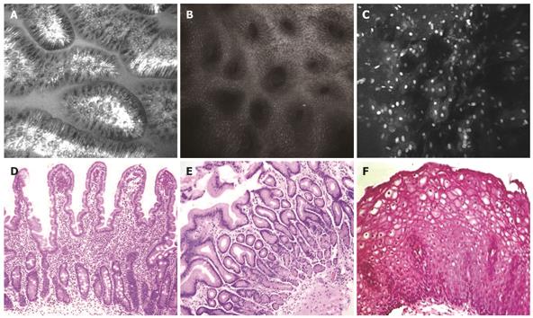
Figure 1 Comparison of confocal images with conventional histological images of the upper GI tract.
A: Confocal image delineating the fine slender fingerlike projections of the duodenal villi; B: Confocal image showing gastric pits; C: Confocal image of non-keratinized squamous epithelium of the esophagus; D: Histological image of duodenum; E: Histological image of gastric antrum; F: Histological image of esophagus.
- Citation: Venkatesh K, Cohen M, Evans C, Delaney P, Thomas S, Taylor C, Abou-Taleb A, Kiesslich R, Thomson M. Feasibility of confocal endomicroscopy in the diagnosis of pediatric gastrointestinal disorders. World J Gastroenterol 2009; 15(18): 2214-2219
- URL: https://www.wjgnet.com/1007-9327/full/v15/i18/2214.htm
- DOI: https://dx.doi.org/10.3748/wjg.15.2214


