Copyright
©2009 The WJG Press and Baishideng.
World J Gastroenterol. May 7, 2009; 15(17): 2139-2144
Published online May 7, 2009. doi: 10.3748/wjg.15.2139
Published online May 7, 2009. doi: 10.3748/wjg.15.2139
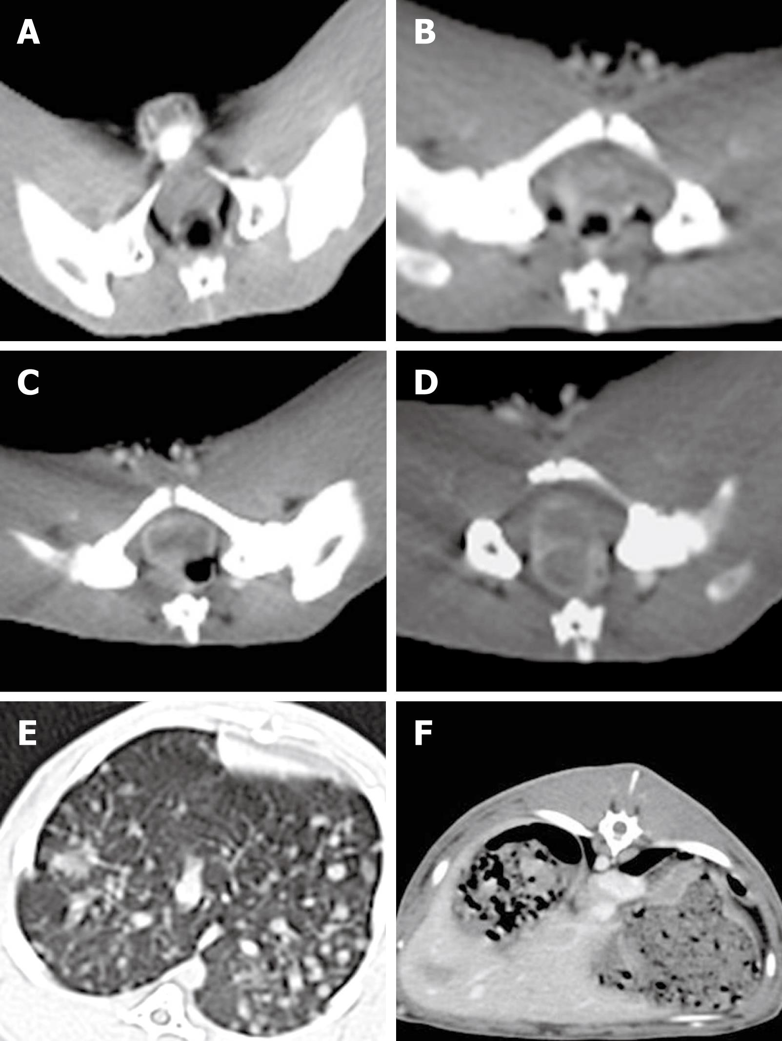
Figure 1 CT enhancement scanning images of rectal wall 2(A), 3(B), 4(C), and 5(D) wk after VX2 cell implantation in the experimental rabbits, and images of metastatic nodes detected in the lung (E) and liver (F), respectively.
- Citation: Liang XM, Tang GY, Cheng YS, Zhou B. Evaluation of a rabbit rectal VX2 carcinoma model using computed tomography and magnetic resonance imaging. World J Gastroenterol 2009; 15(17): 2139-2144
- URL: https://www.wjgnet.com/1007-9327/full/v15/i17/2139.htm
- DOI: https://dx.doi.org/10.3748/wjg.15.2139


