Copyright
©2009 The WJG Press and Baishideng.
World J Gastroenterol. Apr 28, 2009; 15(16): 1951-1957
Published online Apr 28, 2009. doi: 10.3748/wjg.15.1951
Published online Apr 28, 2009. doi: 10.3748/wjg.15.1951
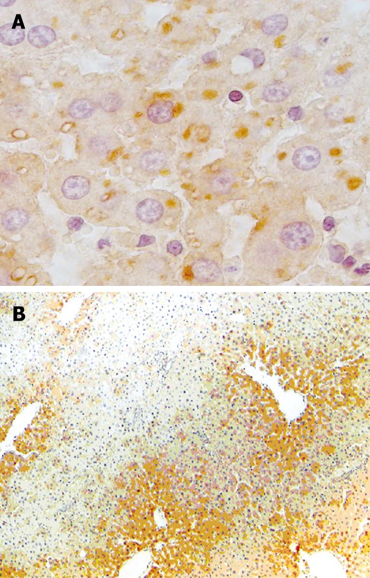
Figure 5 Confirmation of apoptosis by IHC assay in the staining sections of livers exposed to ischemia-reperfusion and the presence of apoptosis cells and/or apoptotic bodies.
A: Representative staining patterns for APAF-1 positive, illustrating the occurrence of apoptosis in the sections of the liver exposed to 60 min ischemia followed by long time of reperfusion; B: APAF-1 positive staining that shows the high level of apoptosis incidence in the pericentral area of the liver exposed to 60 min ischemia followed by long time of reperfusion.
- Citation: Arab HA, Sasani F, Rafiee MH, Fatemi A, Javaheri A. Histological and biochemical alterations in early-stage lobar ischemia-reperfusion in rat liver. World J Gastroenterol 2009; 15(16): 1951-1957
- URL: https://www.wjgnet.com/1007-9327/full/v15/i16/1951.htm
- DOI: https://dx.doi.org/10.3748/wjg.15.1951


