Copyright
©2009 The WJG Press and Baishideng.
World J Gastroenterol. Apr 28, 2009; 15(16): 1951-1957
Published online Apr 28, 2009. doi: 10.3748/wjg.15.1951
Published online Apr 28, 2009. doi: 10.3748/wjg.15.1951
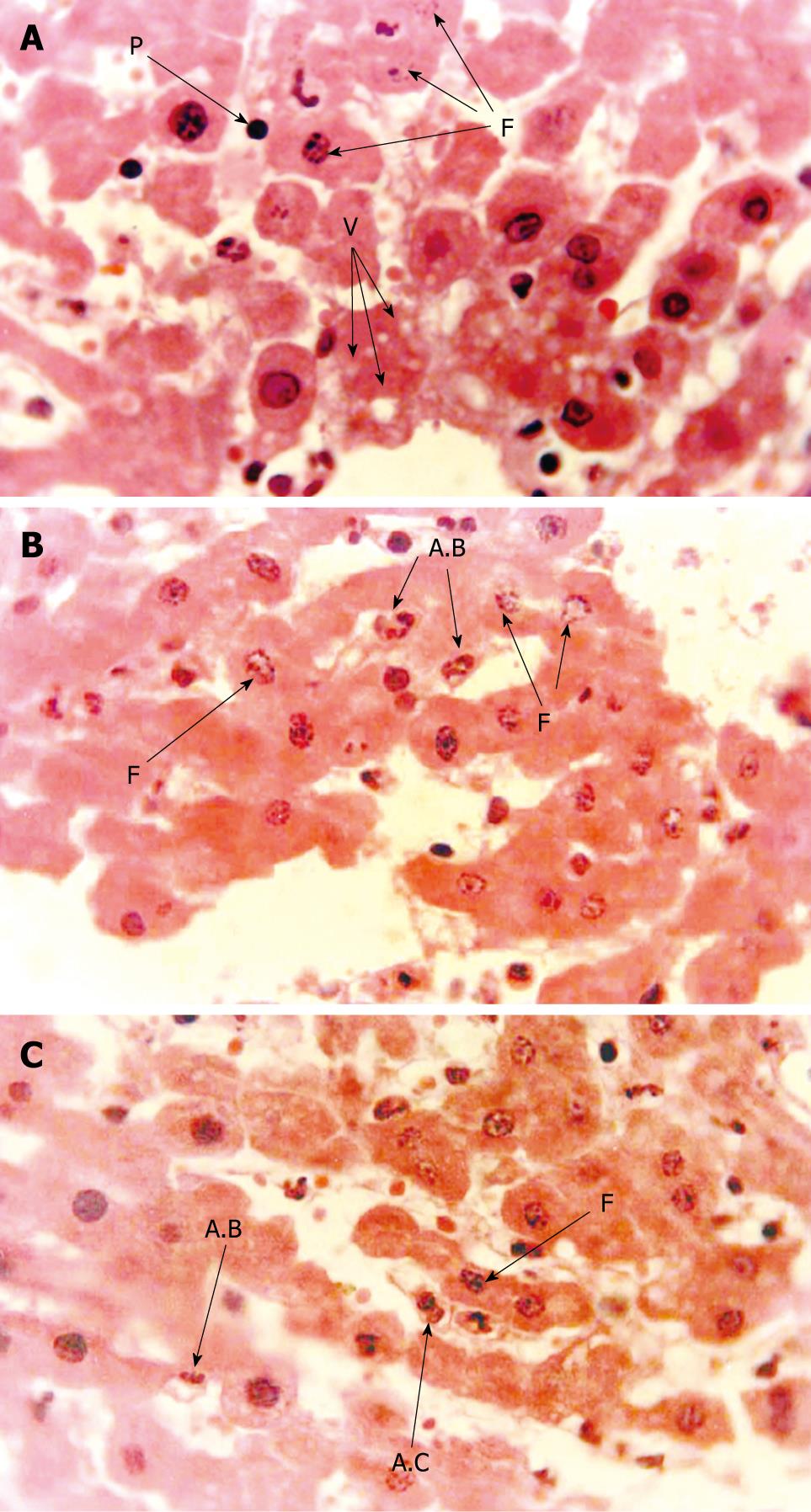
Figure 2 Histological changes in the livers exposed to 60 min lobar ischemia followed by different times of reperfusion.
A: Nuclear pyknosis (P), nuclear fragmentation (F) and cytoplasmic vacuolation in the liver exposed to 60 min ischemia followed by 30 min reperfusion; B: Apoptotic bodies (A.B) and nuclear fragmentation (F) in the liver exposed to 60 min ischemia followed by 60 min reperfusion; C: Nuclear fragmentation (F), apoptotic cell (A.C) and apoptotic bodies (A.B) in the liver exposed to 60 min ischemia followed by120 min reperfusion.
- Citation: Arab HA, Sasani F, Rafiee MH, Fatemi A, Javaheri A. Histological and biochemical alterations in early-stage lobar ischemia-reperfusion in rat liver. World J Gastroenterol 2009; 15(16): 1951-1957
- URL: https://www.wjgnet.com/1007-9327/full/v15/i16/1951.htm
- DOI: https://dx.doi.org/10.3748/wjg.15.1951


