Copyright
©2009 The WJG Press and Baishideng.
World J Gastroenterol. Apr 21, 2009; 15(15): 1901-1903
Published online Apr 21, 2009. doi: 10.3748/wjg.15.1901
Published online Apr 21, 2009. doi: 10.3748/wjg.15.1901
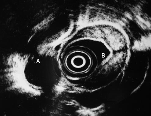
Figure 3 Endoscopic ultrasonography shows a transmural thickening of the esophageal wall and a heterogeneous, mainly hyperechoic, submucosal lesion.
A: Thoracic aorta, B: Water-filled balloon, T: Tumor.
- Citation: Kalogeropoulos IV, Chalazonitis AN, Tsolaki S, Laspas F, Ptohis N, Neofytou I, Rontogianni D. A case of primary isolated non-Hodgkin’s lymphoma of the esophagus in an immunocompetent patient. World J Gastroenterol 2009; 15(15): 1901-1903
- URL: https://www.wjgnet.com/1007-9327/full/v15/i15/1901.htm
- DOI: https://dx.doi.org/10.3748/wjg.15.1901


