Copyright
©2009 The WJG Press and Baishideng.
World J Gastroenterol. Apr 21, 2009; 15(15): 1836-1842
Published online Apr 21, 2009. doi: 10.3748/wjg.15.1836
Published online Apr 21, 2009. doi: 10.3748/wjg.15.1836
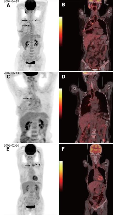
Figure 2 A 62-year-women had esophageal cancer resection.
She underwent three PET/CT scans (1.5, 4.5 and 11 mo after treatment, respectively) as part of routine post-operative surveillance. The first PET/CT imaging (1.5 mo) revealed hypermetabolic activity in the anastomosis and supraclavicular lymph nodes (arrows, A, B). The second PET/CT imaging (4.5 mo) showed decreasing of hypermetabolic activity at anastomosis and supraclavicular lymph nodes (arrows, C, D). The third PET/CT imaging (11 mo) showed no abnormal FDG uptake at anastomosis, but revealed new focal hypermetabolic activity at left supraclavicular lymph nodes (arrows, E, F). Inflammation of lymph nodes and anastomosis at the first and second PET/CT scan were confirmed by the third PET/CT examination. New relapse at the left supraclavicular lymph nodes was later verified by biopsy.
- Citation: Sun L, Su XH, Guan YS, Pan WM, Luo ZM, Wei JH, Zhao L, Wu H. Clinical usefulness of 18F-FDG PET/CT in the restaging of esophageal cancer after surgical resection and radiotherapy. World J Gastroenterol 2009; 15(15): 1836-1842
- URL: https://www.wjgnet.com/1007-9327/full/v15/i15/1836.htm
- DOI: https://dx.doi.org/10.3748/wjg.15.1836


