Copyright
©2009 The WJG Press and Baishideng.
World J Gastroenterol. Apr 14, 2009; 15(14): 1782-1785
Published online Apr 14, 2009. doi: 10.3748/wjg.15.1782
Published online Apr 14, 2009. doi: 10.3748/wjg.15.1782
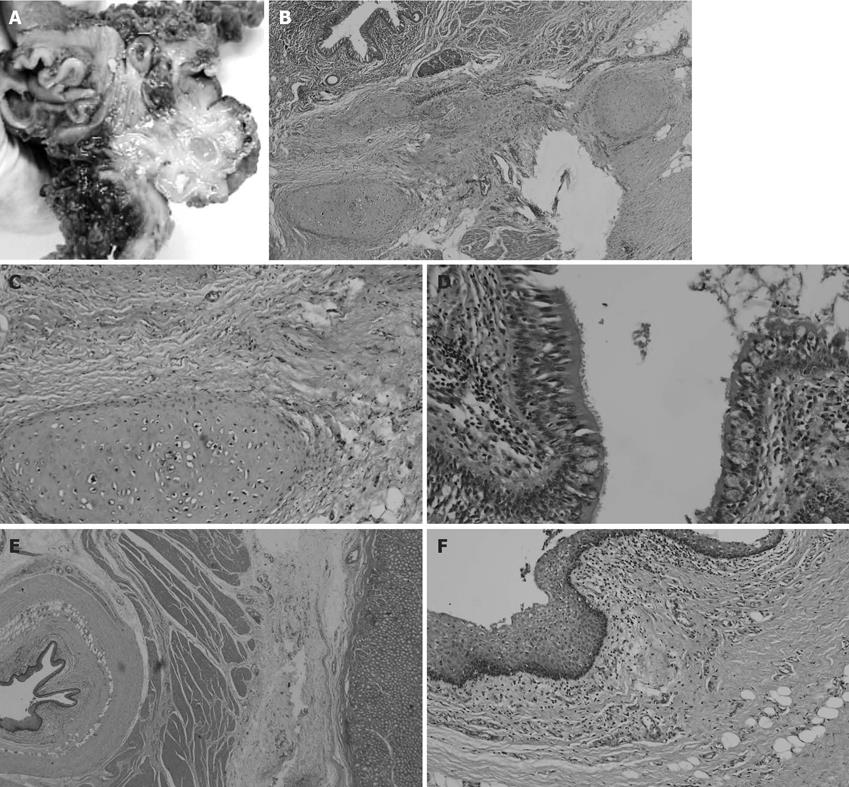
Figure 3 GT was pathologically examined under macroscopy and microscopy.
A: The mass was about 5.0 cm × 5.3 cm × 2.3 cm, and it derived from the inferior wall of the cardiac orifice; B: Photomicrograph of an area composed respiratory epithelium and cartilage without immature teratoma components (HE, × 20); C, D: The cartilage (C) and the respiratory epithelium (D) were amplified (HE, × 100); E: The three layers, mucous layer, submucous membrane, and muscular layer, of the wall of the stomach were clear. The tissue of GT was derived from the muscular layer of the stomach (HE, × 20); F: The Squamous cell was amplified (HE, × 100). C, D: The cartilage (C) and the respiratory epithelium (D) were amplified (HE, × 100); E: The three layers, mucous layer, submucous membrane, and muscular layer, of the wall of the stomach were clear. The tissue of GT was derived from the muscular layer of the stomach (HE, × 20); F: The Squamous cell was amplified (HE, × 100).
- Citation: Liu L, Zhuang W, Chen Z, Zhou Y, Huang XR. Primary gastric teratoma on the cardiac orifice in an adult. World J Gastroenterol 2009; 15(14): 1782-1785
- URL: https://www.wjgnet.com/1007-9327/full/v15/i14/1782.htm
- DOI: https://dx.doi.org/10.3748/wjg.15.1782


