Copyright
©2009 The WJG Press and Baishideng.
World J Gastroenterol. Apr 14, 2009; 15(14): 1738-1743
Published online Apr 14, 2009. doi: 10.3748/wjg.15.1738
Published online Apr 14, 2009. doi: 10.3748/wjg.15.1738
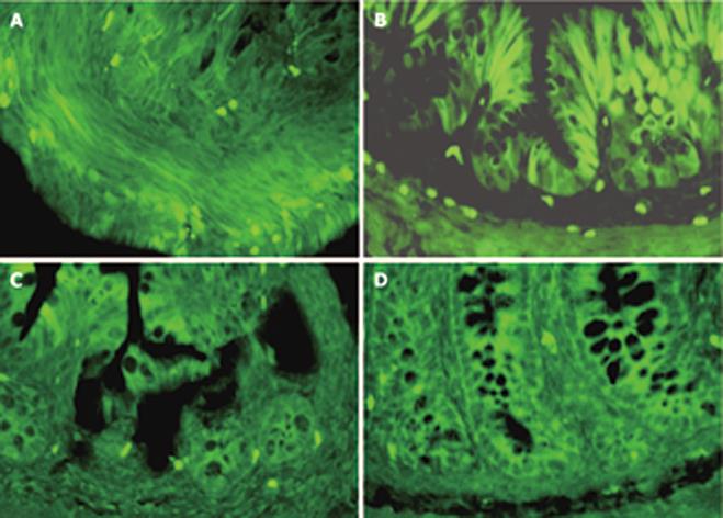
Figure 3 Immunofluorescence localization of AFP in the developing rat colons.
A: In e18.5, AFP positive staining can be detected in the epithelium and mesenchymal tissues; B: At P0, positive cells were located at the base of the crypts and scattered on the epithelium; C, D: Only a few positive cells restricted to the base of the crypts between 14 and 21 d, and no positive cells can be detected in adult rat colons. (× 200). C, D: Only a few positive cells restricted to the base of the crypts between 14 and 21 d, and no positive cells can be detected in adult rat colons. (× 200).
- Citation: Liu XY, Dong D, Sun P, Du J, Gu L, Ge YB. Expression and location of α-fetoprotein during rat colon development. World J Gastroenterol 2009; 15(14): 1738-1743
- URL: https://www.wjgnet.com/1007-9327/full/v15/i14/1738.htm
- DOI: https://dx.doi.org/10.3748/wjg.15.1738


