Copyright
©2009 The WJG Press and Baishideng.
World J Gastroenterol. Apr 14, 2009; 15(14): 1708-1718
Published online Apr 14, 2009. doi: 10.3748/wjg.15.1708
Published online Apr 14, 2009. doi: 10.3748/wjg.15.1708
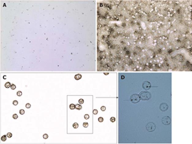
Figure 4 Images documenting LCM of carbon-labeled Kupffer cells.
A: Static image of carbon-labeled liver section overlaid with xylene (10 × magnification); B: Same tissue section after the evaporation of xylene and capture of carbon-labeled cells (10 ×); C: Captured cells visualized on the CapSure LCM cap (20 ×); D: Same cap mounted with water and a coverslip showing carbon within microdissected cells (arrows, 40 ×).
- Citation: Gehring S, Sabo E, Martin MES, Dickson EM, Cheng CW, Gregory SH. Laser capture microdissection and genetic analysis of carbon-labeled Kupffer cells. World J Gastroenterol 2009; 15(14): 1708-1718
- URL: https://www.wjgnet.com/1007-9327/full/v15/i14/1708.htm
- DOI: https://dx.doi.org/10.3748/wjg.15.1708


