Copyright
©2009 The WJG Press and Baishideng.
World J Gastroenterol. Apr 7, 2009; 15(13): 1656-1659
Published online Apr 7, 2009. doi: 10.3748/wjg.15.1656
Published online Apr 7, 2009. doi: 10.3748/wjg.15.1656
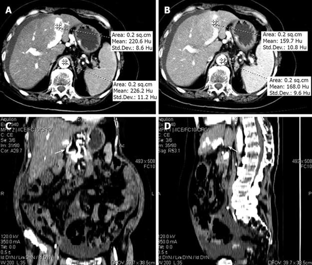
Figure 3 Contrast-enhanced CT showing a LPV density of 220.
6 Hu and an abdominal aorta density of 226.2 Hu at artery phase (A), a LPV density of 159.7 Hu and an abdominal aorta density of 168.0 Hu at portal venous phase (B), and CT of coronal section (C) and sagittal section (D) showing the direct shunt of APF.
- Citation: Lu ZY, Ao JY, Jiang TA, Peng ZY, Wang ZK. A large congenital and solitary intrahepatic arterioportal fistula in an old woman. World J Gastroenterol 2009; 15(13): 1656-1659
- URL: https://www.wjgnet.com/1007-9327/full/v15/i13/1656.htm
- DOI: https://dx.doi.org/10.3748/wjg.15.1656


