Copyright
©2009 The WJG Press and Baishideng.
World J Gastroenterol. Mar 28, 2009; 15(12): 1431-1442
Published online Mar 28, 2009. doi: 10.3748/wjg.15.1431
Published online Mar 28, 2009. doi: 10.3748/wjg.15.1431
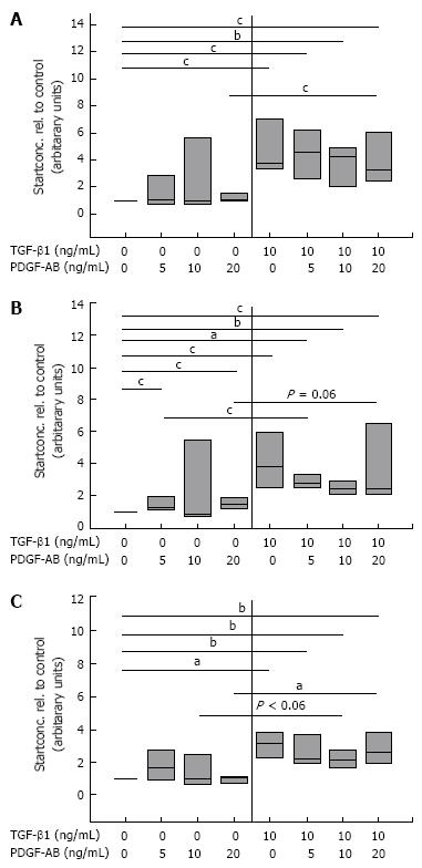
Figure 6 Quantitative mRNA analyses of FN (A) and the splicing forms FN ED-A (B), and FN ED-B (C) by real-time PCR in CLPF treated with TGF-β1.
Control CLPF (n = 7) were pre-incubated with and without 10 ng/mL TGF-β1 for 6 d, the monolayer wounded with a comb, and incubated for further 4 h with increasing concentrations of PDGF-AB in conditioned medium. mRNA was isolated and cDNA transcribed. cDNA start concentration of the untreated control (0 ng/mL TGF-β1 and 0 ng/mL PDGF-AB) was set as 1. Paired t-test: aP < 0.005; bP < 0.01; cP < 0.05.
- Citation: Brenmoehl J, Miller SN, Hofmann C, Vogl D, Falk W, Schölmerich J, Rogler G. Transforming growth factor-β1 induces intestinal myofibroblast differentiation and modulates their migration. World J Gastroenterol 2009; 15(12): 1431-1442
- URL: https://www.wjgnet.com/1007-9327/full/v15/i12/1431.htm
- DOI: https://dx.doi.org/10.3748/wjg.15.1431


