Copyright
©2009 The WJG Press and Baishideng.
World J Gastroenterol. Mar 21, 2009; 15(11): 1381-1387
Published online Mar 21, 2009. doi: 10.3748/wjg.15.1381
Published online Mar 21, 2009. doi: 10.3748/wjg.15.1381
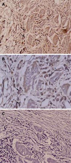
Figure 2 Immunohistochemical staining of p53 protein in diffused type-poorly differentiated gastric cancer.
p53 protein over-expression is seen primarily on cell nucleus. A: Positive nuclear staining (× 200); B: Positive nuclear staining (× 400); C: Negative control (× 200).
-
Citation: Karim S, Ali A. Correlation of p53 over-expression and alteration in
p53 gene detected by polymerase chain reaction-single strand conformation polymorphism in adenocarcinoma of gastric cancer patients from India. World J Gastroenterol 2009; 15(11): 1381-1387 - URL: https://www.wjgnet.com/1007-9327/full/v15/i11/1381.htm
- DOI: https://dx.doi.org/10.3748/wjg.15.1381


