Copyright
©2009 The WJG Press and Baishideng.
World J Gastroenterol. Mar 21, 2009; 15(11): 1373-1380
Published online Mar 21, 2009. doi: 10.3748/wjg.15.1373
Published online Mar 21, 2009. doi: 10.3748/wjg.15.1373
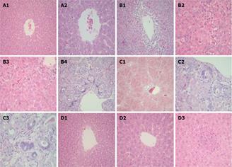
Figure 2 Photomicrographs of liver specimens stained with H&E.
A: Liver from control rat showing normal liver histology with unremarkable central vein (A1, × 20 and A2, × 40); B: Liver from rat treated with DENA showing central vein surrounded by extensive necrosis and inflammatory infiltrate (B1, × 20), considerable hepatocyte necrosis represented with arrows (B2 and B3, × 40) and portal tract with bile duct proliferation and marked atypia (B4, × 20); C: Liver from rat treated with DENA plus D-carnitine-mildronate showing diffuse bridging fibrosis and nodule formation (C1, × 10) and bile ducts with marked reactive atypia showing nuclear enlargement, high nuclear/cytoplasmic ratio and prominent nucleoli (C2 and C3, × 40); D: Liver from rat treated with DENA and L-carnitine showing normal liver (D1, × 20) with unremarkable central vein (D2, × 40) and hepatic lobule with a focus of inflammatory infiltrate but no necrosis (D3, × 40).
- Citation: Al-Rejaie SS, Aleisa AM, Al-Yahya AA, Bakheet SA, Alsheikh A, Fatani AG, Al-Shabanah OA, Sayed-Ahmed MM. Progression of diethylnitrosamine-induced hepatic carcinogenesis in carnitine-depleted rats. World J Gastroenterol 2009; 15(11): 1373-1380
- URL: https://www.wjgnet.com/1007-9327/full/v15/i11/1373.htm
- DOI: https://dx.doi.org/10.3748/wjg.15.1373


