Copyright
©2009 The WJG Press and Baishideng.
World J Gastroenterol. Mar 21, 2009; 15(11): 1359-1366
Published online Mar 21, 2009. doi: 10.3748/wjg.15.1359
Published online Mar 21, 2009. doi: 10.3748/wjg.15.1359
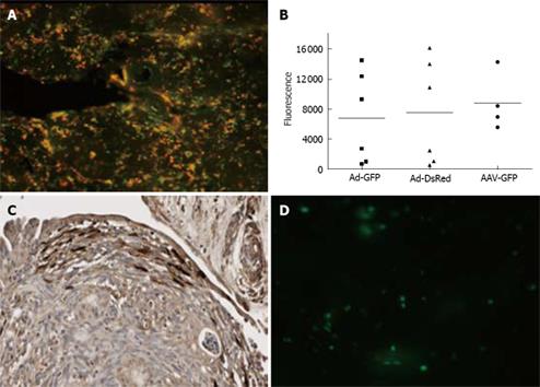
Figure 5 Transduction of pancreatic tissue slices with different viral vectors.
A: Normal human pancreatic tissue slices are incubated with 1.0 × 10⁸ Ad5.GFP and 1.0 × 10⁸ Ad5.dsRed. After 48 h presence of GFP and ds.Red was detected using a fluorescent microscope; B: Normal pancreas slices were infected with 1.0 × 10⁸ genomic copies per slice of Ad5.GFP and Ad5.dsRED, or 1.2 × 10¹⁰ viral genomes AAV2-GFP. At 48 h after infection slices were lysed, GFP and ds.Red were quantified using the Novostar; C: Pancreatic adenocarcinoma explants were transduced with Ad-GFP. At 72 h after virus addition expression of GFP was detected using an anti-GFP antibody and counterstained with hematoxylin; D: GFP expression in normal pancreas at 72 h after incubation with lentivirus-GFP.
-
Citation: Geer MAV, Kuhlmann KF, Bakker CT, Kate FJT, Elferink RPO, Bosma PJ.
Ex-vivo evaluation of gene therapy vectors in human pancreatic (cancer) tissue slices. World J Gastroenterol 2009; 15(11): 1359-1366 - URL: https://www.wjgnet.com/1007-9327/full/v15/i11/1359.htm
- DOI: https://dx.doi.org/10.3748/wjg.15.1359


