Copyright
©2009 The WJG Press and Baishideng.
World J Gastroenterol. Mar 21, 2009; 15(11): 1289-1300
Published online Mar 21, 2009. doi: 10.3748/wjg.15.1289
Published online Mar 21, 2009. doi: 10.3748/wjg.15.1289
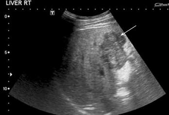
Figure 1 HCC.
B-mode US demonstrates a heterogeneous hypoechoic solid liver lesion (arrow) in segment IV, confirmed to be HCC at histological examination. Note the background of an echogenic liver with an irregular surface.
- Citation: Ariff B, Lloyd CR, Khan S, Shariff M, Thillainayagam AV, Bansi DS, Khan SA, Taylor-Robinson SD, Lim AK. Imaging of liver cancer. World J Gastroenterol 2009; 15(11): 1289-1300
- URL: https://www.wjgnet.com/1007-9327/full/v15/i11/1289.htm
- DOI: https://dx.doi.org/10.3748/wjg.15.1289


