Copyright
©2009 The WJG Press and Baishideng.
World J Gastroenterol. Mar 14, 2009; 15(10): 1246-1253
Published online Mar 14, 2009. doi: 10.3748/wjg.15.1246
Published online Mar 14, 2009. doi: 10.3748/wjg.15.1246
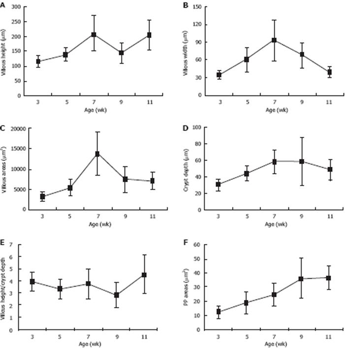
Figure 2 Morphological analysis of villous-crypt axis: All morphological parameters matured as age increased.
The results were presented as mean ± SD from 5 rats. A: Villous height increased at 3 wk postpartum, decreased 7 to 9 wk postpartum, and increased again after 9 wk postpartum; B: Villous width increased at 3 wk postpartum, peaked at 7 wk postpartum; C: Villous area increased significantly between 5 and 7 wk postpartum, peaked at 7 wk postpartum; D: Crypt depth increased from 3 to 7 wk postpartum and decreased slightly at 9 wk postpartum; E: Ratio of villous height to crypt depth were relatively stable and increased significantly from 9 to 11 wk postpartum; F: PP increased from 3 to 11 wk postpartum.
- Citation: Zhou YJ, Gao J, Yang HM, Zhu JX, Chen TX, He ZJ. Morphology and ontogeny of dendritic cells in rats at different development periods. World J Gastroenterol 2009; 15(10): 1246-1253
- URL: https://www.wjgnet.com/1007-9327/full/v15/i10/1246.htm
- DOI: https://dx.doi.org/10.3748/wjg.15.1246


