Copyright
©2008 The WJG Press and Baishideng.
World J Gastroenterol. Mar 7, 2008; 14(9): 1463-1466
Published online Mar 7, 2008. doi: 10.3748/wjg.14.1463
Published online Mar 7, 2008. doi: 10.3748/wjg.14.1463
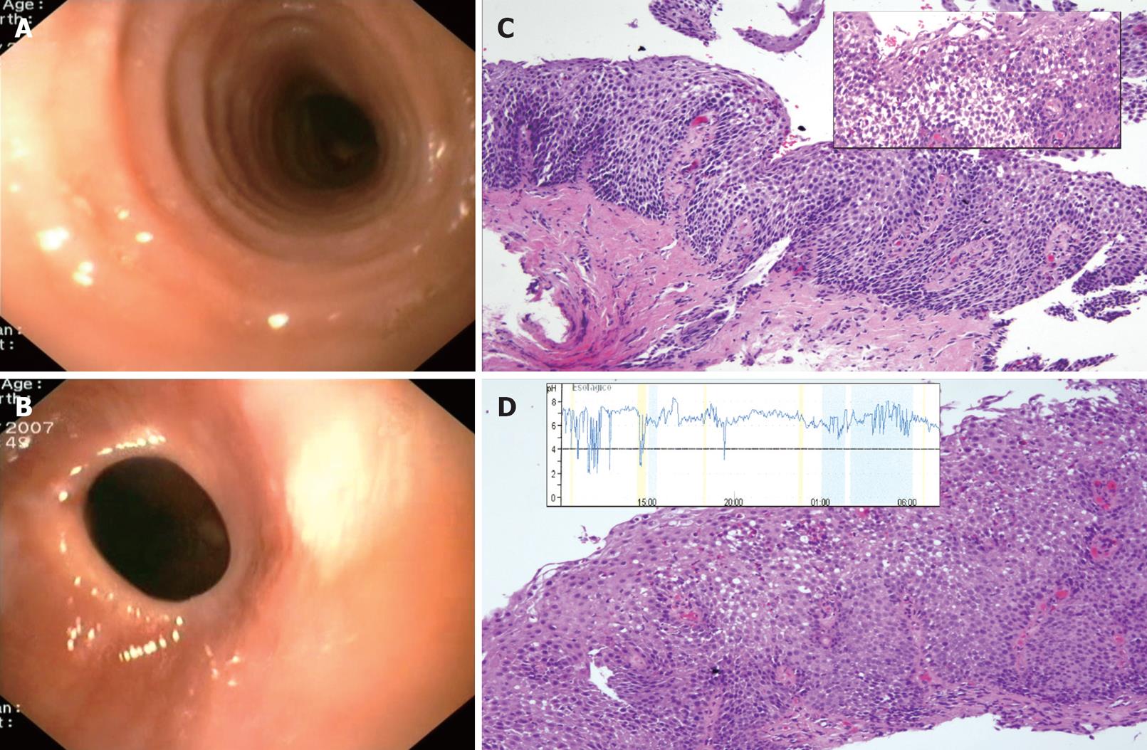
Figure 2 Case 2.
A: Endoscopic view of the upper esophagus showing multiple concentric rings resembling mucosal undulations; B: Endoscopic picture of a peptic stricture at cardias 2 mo after dilation, with gentle pressure of the endoscope; C: Biopsies of a multiringed esophagus demonstrating severe lamina propia fibrosis at the bottom of the image, as well as marked papillae elongation and basal zone hyperplasia (HE, × 100). In the upper right box, intense intercellular edema, basal zone hyperplasia and dense eosinophilic infiltrate (37eo/ HPF, × 400) towards the surface strata (HE, × 200); D: Persistent histopathologic features (32 eo/HPF, × 400) in spite of effective PPI therapy, as shown by 24 h pH esophageal monitoring (box).
- Citation: Molina-Infante J, Ferrando-Lamana L, Mateos-Rodríguez JM, Pérez-Gallardo B, Prieto-Bermejo AB. Overlap of reflux and eosinophilic esophagitis in two patients requiring different therapies: A review of the literature. World J Gastroenterol 2008; 14(9): 1463-1466
- URL: https://www.wjgnet.com/1007-9327/full/v14/i9/1463.htm
- DOI: https://dx.doi.org/10.3748/wjg.14.1463


