Copyright
©2008 The WJG Press and Baishideng.
World J Gastroenterol. Mar 7, 2008; 14(9): 1463-1466
Published online Mar 7, 2008. doi: 10.3748/wjg.14.1463
Published online Mar 7, 2008. doi: 10.3748/wjg.14.1463
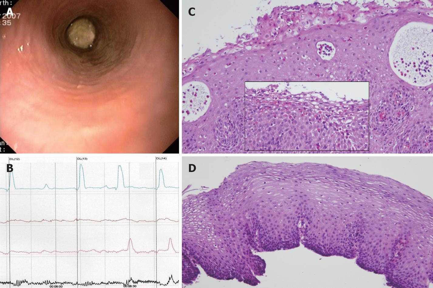
Figure 1 Case 1.
A: Emergency endoscopy showing a meat bolus impacted at mid-distal esophagus with normal mucosa; B: Esophageal manometry demonstrated that two thirds of all contractions in the distal esophagus were simultaneous or interrupted contraction waves; C: Prominent eosinophil microabscesses in upper-mid esophagus biopsies (HE, × 100). In the box and on top of the image, a dense eosinophilic infiltrate (31eo/HPF × 400) predominantly spread over the superficial layers can be observed (HE, × 200); D: Normal upper-mid squamous epithelium after PPI therapy (HE, × 100).
- Citation: Molina-Infante J, Ferrando-Lamana L, Mateos-Rodríguez JM, Pérez-Gallardo B, Prieto-Bermejo AB. Overlap of reflux and eosinophilic esophagitis in two patients requiring different therapies: A review of the literature. World J Gastroenterol 2008; 14(9): 1463-1466
- URL: https://www.wjgnet.com/1007-9327/full/v14/i9/1463.htm
- DOI: https://dx.doi.org/10.3748/wjg.14.1463


