Copyright
©2008 The WJG Press and Baishideng.
World J Gastroenterol. Feb 28, 2008; 14(8): 1212-1217
Published online Feb 28, 2008. doi: 10.3748/wjg.14.1212
Published online Feb 28, 2008. doi: 10.3748/wjg.14.1212
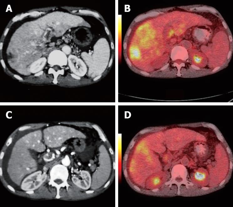
Figure 5 Contrast CT scan showing a diffuse tumor and portal vein thrombus in a 44-year-old man.
During the arterial phase, the thrombus appears as a hypodense intraluminal area with mild enhancement at the LV (white arrows, A) and PV (white arrows, C). PET/CT fused images reveal a highly metabolic mass and a highly metabolic thrombus at the LV and PV, respectively (white arrows, B, D).
- Citation: Sun L, Guan YS, Pan WM, Chen GB, Luo ZM, Wei JH, Wu H. Highly metabolic thrombus of the portal vein: 18F fluorodeoxyglucose positron emission tomography/computer tomography demonstration and clinical significance in hepatocellular carcinoma. World J Gastroenterol 2008; 14(8): 1212-1217
- URL: https://www.wjgnet.com/1007-9327/full/v14/i8/1212.htm
- DOI: https://dx.doi.org/10.3748/wjg.14.1212


